
















©2009 The WJG Press and Baishideng.
World J Gastroenterol. Feb 28, 2009; 15(8): 1010-1013
Published online Feb 28, 2009. doi: 10.3748/wjg.15.1010
Published online Feb 28, 2009. doi: 10.3748/wjg.15.1010
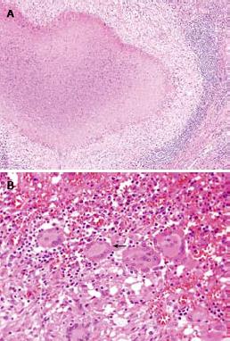
Figure 4 Histological image of resected specimen.
A: Photograph showing the epithelioid granuloma with central caseous necrosis (HE, original magnification, × 100); B: multinucleated Langhans’ giant cell with surrounding lymphocytes (arrow) (HE, original magnification, × 400).
- Citation: Hong SG, Kim JS, Joo MK, Lee KG, Kim KH, Oh CR, Park JJ, Bak YT. Pancreatic tuberculosis masquerading as pancreatic serous cystadenoma. World J Gastroenterol 2009; 15(8): 1010-1013
- URL: https://www.wjgnet.com/1007-9327/full/v15/i8/1010.htm
- DOI: https://dx.doi.org/10.3748/wjg.15.1010