
















©2009 The WJG Press and Baishideng.
World J Gastroenterol. Dec 28, 2009; 15(48): 6129-6133
Published online Dec 28, 2009. doi: 10.3748/wjg.15.6129
Published online Dec 28, 2009. doi: 10.3748/wjg.15.6129
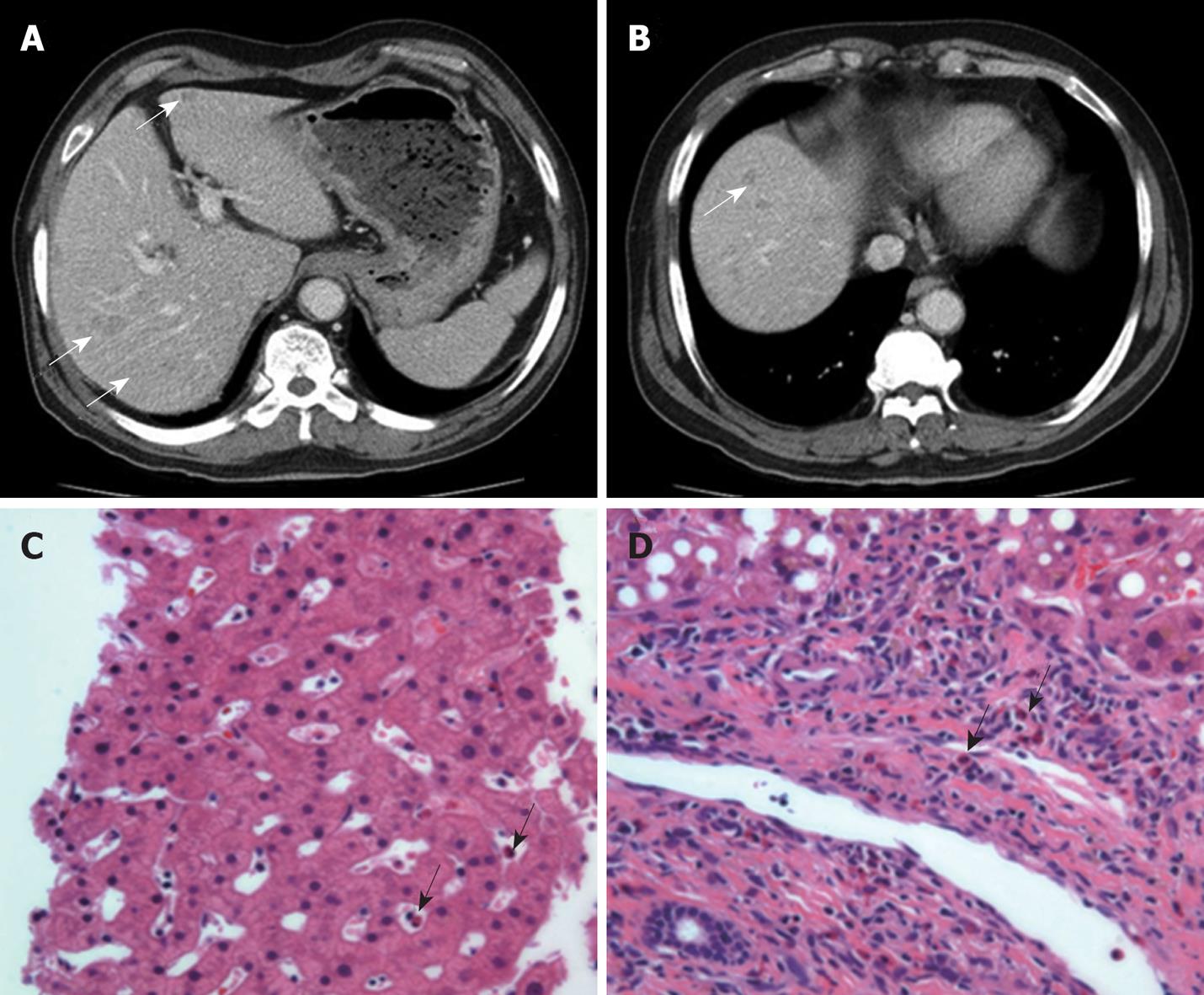
Figure 3 Abdominal-pelvic CT scan and liver biopsy findings.
A, B: There were multiple, small, ill-defined low densities found in the liver (white arrows); C, D: Microscopically, eosinophilic infiltration was noted in the sinusoid and the portal tract (black arrows) (HE stain, × 400).
- Citation: Park TY, Choi CH, Yang SY, Oh IS, Song ID, Lee HW, Kim HJ, Do JH, Chang SK, Cho AR, Cha YJ. A case of hypereosinophilic syndrome presenting with intractable gastric ulcers. World J Gastroenterol 2009; 15(48): 6129-6133
- URL: https://www.wjgnet.com/1007-9327/full/v15/i48/6129.htm
- DOI: https://dx.doi.org/10.3748/wjg.15.6129