
















©2009 The WJG Press and Baishideng.
World J Gastroenterol. Dec 28, 2009; 15(48): 6044-6051
Published online Dec 28, 2009. doi: 10.3748/wjg.15.6044
Published online Dec 28, 2009. doi: 10.3748/wjg.15.6044
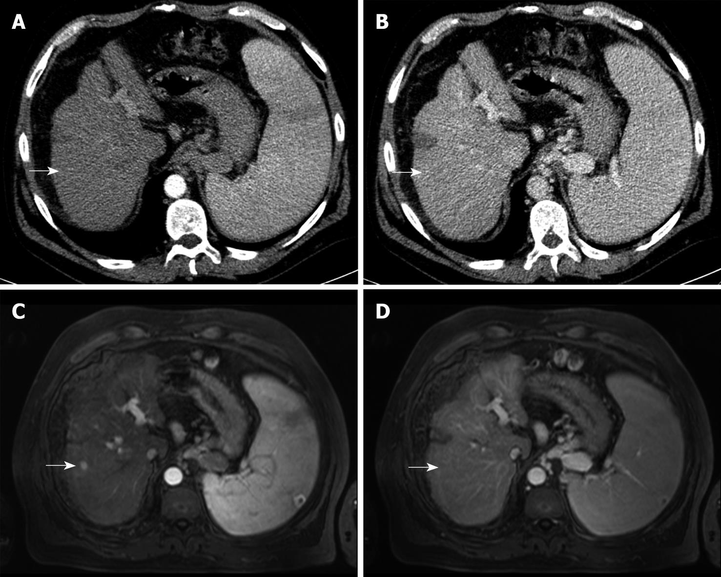
Figure 2 70-year-old man with biopsy-proven HCC.
Detection of an additional tumour nodule by MRI, size 10 mm (size category ≤ 10 mm). MDCT does not show any contrast enhancement in the arterial (A, arrow) or portal venous phase (B, arrow). MRI arterial phase depicts one more tumour nodule (C, arrow) which is hypo- to isointense on the portal venous phase (D, arrow).
-
Citation: Pitton MB, Kloeckner R, Herber S, Otto G, Kreitner KF, Dueber C. MRI
versus 64-row MDCT for diagnosis of hepatocellular carcinoma. World J Gastroenterol 2009; 15(48): 6044-6051 - URL: https://www.wjgnet.com/1007-9327/full/v15/i48/6044.htm
- DOI: https://dx.doi.org/10.3748/wjg.15.6044