
















©2009 The WJG Press and Baishideng.
World J Gastroenterol. Sep 21, 2009; 15(35): 4467-4470
Published online Sep 21, 2009. doi: 10.3748/wjg.15.4467
Published online Sep 21, 2009. doi: 10.3748/wjg.15.4467
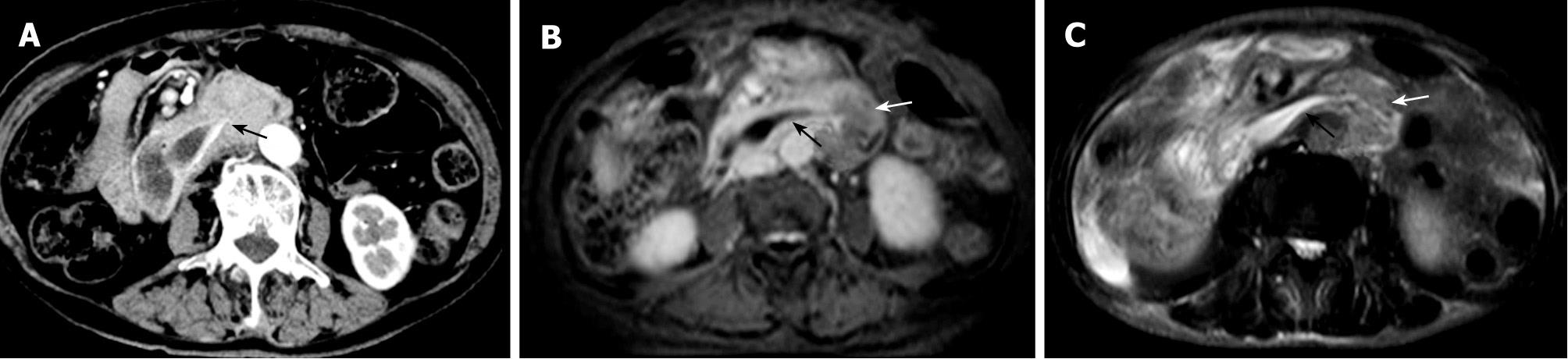
Figure 2 Anomalous termination of the common bile duct.
A: Axial image from CT scan shows the termination of the common bile duct at the distal portion of the horizontal part of the duodenum (arrow); B and C: MR images demonstrate a tapered configuration of the common bile duct (black arrows) and a mass located on the proximal jejunum (white arrows). (These photographs were processed by Adobe Photoshop Creative Suite 2.).
- Citation: Wu DS, Chen WX, Wang XP. Ectopic pancreaticobiliary drainage accompanied by proximal jejunal adenoma: A case report. World J Gastroenterol 2009; 15(35): 4467-4470
- URL: https://www.wjgnet.com/1007-9327/full/v15/i35/4467.htm
- DOI: https://dx.doi.org/10.3748/wjg.15.4467