
















©2009 The WJG Press and Baishideng.
World J Gastroenterol. Sep 21, 2009; 15(35): 4434-4438
Published online Sep 21, 2009. doi: 10.3748/wjg.15.4434
Published online Sep 21, 2009. doi: 10.3748/wjg.15.4434
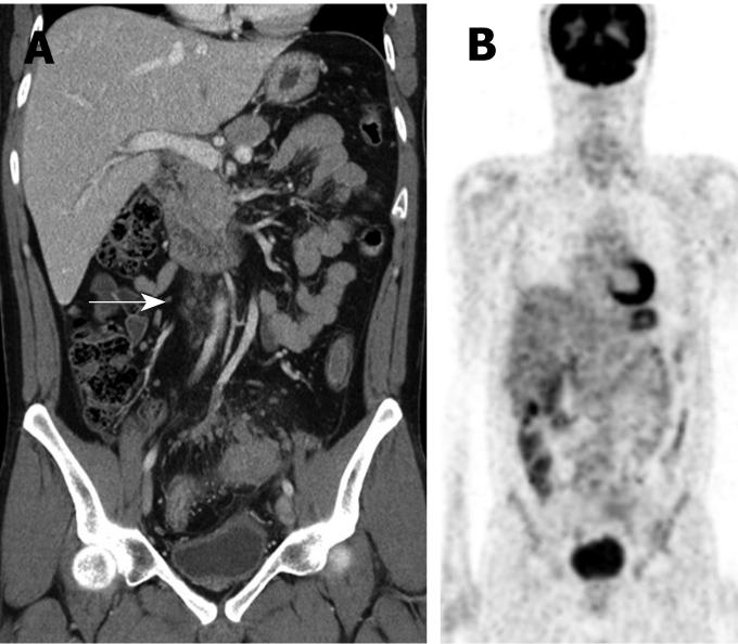
Figure 2 False positive metastatic PAN on CT.
A 38-year-old man with colon cancer underwent CT and PET. (A) Coronal CT scan shows multiple enlarged (short diameter was 8 mm in the largest one) PAN (arrow), but (B) coronal view of PET image shows negative fluorodeoxyglucose uptake in these lymph nodes. This patient had underlying inflammatory bowel disease of ulcerative colitis.
- Citation: Lee MJ, Yun MJ, Park MS, Cha SH, Kim MJ, Lee JD, Kim KW. Paraaortic lymph node metastasis in patients with intra-abdominal malignancies: CT vs PET. World J Gastroenterol 2009; 15(35): 4434-4438
- URL: https://www.wjgnet.com/1007-9327/full/v15/i35/4434.htm
- DOI: https://dx.doi.org/10.3748/wjg.15.4434