
















©2009 The WJG Press and Baishideng.
World J Gastroenterol. Sep 21, 2009; 15(35): 4434-4438
Published online Sep 21, 2009. doi: 10.3748/wjg.15.4434
Published online Sep 21, 2009. doi: 10.3748/wjg.15.4434
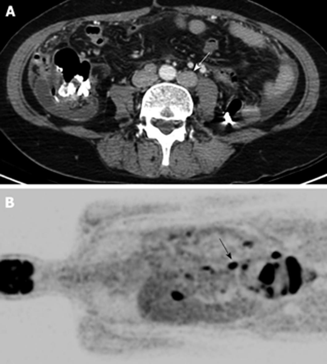
Figure 1 Typical case of metastatic paraaortic lymph node (PAN) on both computed tomography (CT) and positron emission tomography (PET).
A 52-year-old woman with ovarian cancer underwent CT and PET. (A) Axial CT scan shows enlarged (short diameter was 16 mm) and conglomerated PAN (arrow) and (B) coronal view of PET image shows positive FDG uptake in this PAN (arrow).
- Citation: Lee MJ, Yun MJ, Park MS, Cha SH, Kim MJ, Lee JD, Kim KW. Paraaortic lymph node metastasis in patients with intra-abdominal malignancies: CT vs PET. World J Gastroenterol 2009; 15(35): 4434-4438
- URL: https://www.wjgnet.com/1007-9327/full/v15/i35/4434.htm
- DOI: https://dx.doi.org/10.3748/wjg.15.4434