
















©2009 The WJG Press and Baishideng.
World J Gastroenterol. Aug 28, 2009; 15(32): 4070-4074
Published online Aug 28, 2009. doi: 10.3748/wjg.15.4070
Published online Aug 28, 2009. doi: 10.3748/wjg.15.4070
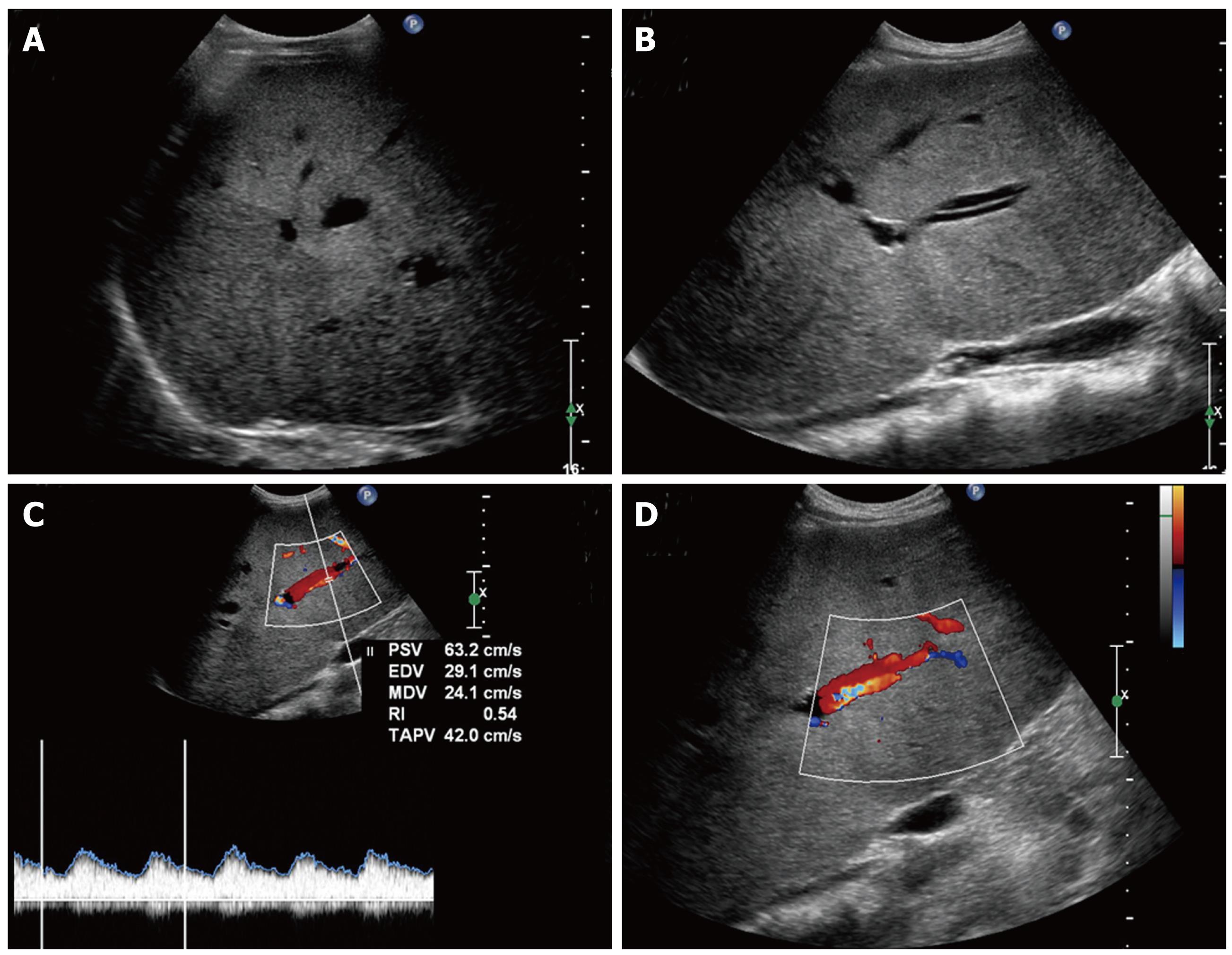
Figure 1 US scan showing a large hypoechoic area compared to surrounding parenchyma (A) and the image of “parallel channel” sign (B).
Color-Doppler demonstrates the “pseudoparallel channel” sign, characterized by dilated intrahepatic arterial branch with an adjacent portal venous tract, and the low hepatic artery RI (C, D).
- Citation: Tenca A, Massironi S, Colli A, Basilisco G, Conte D. “Pseudotumoral” hepatic pattern in acute alcoholic hepatitis: A case report. World J Gastroenterol 2009; 15(32): 4070-4074
- URL: https://www.wjgnet.com/1007-9327/full/v15/i32/4070.htm
- DOI: https://dx.doi.org/10.3748/wjg.15.4070