
















©2009 The WJG Press and Baishideng.
World J Gastroenterol. Aug 28, 2009; 15(32): 4049-4054
Published online Aug 28, 2009. doi: 10.3748/wjg.15.4049
Published online Aug 28, 2009. doi: 10.3748/wjg.15.4049
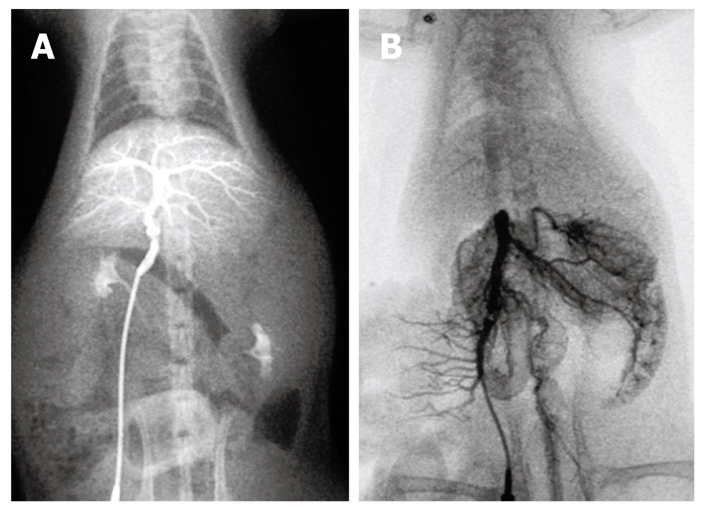
Figure 4 Portal venography in control rat.
A: The intrahepatic portal vein was shown as tree twig branches. There was little contrast medium in the mesenteric vein and the splenic vein; B: After ligation of the portal vein, images of the superior and inferior mesenteric vein and the splenic vein appeared simultaneously, but no collateral circulation could be seen.
- Citation: Wen Z, Zhang JZ, Xia HM, Yang CX, Chen YJ. Stability of a rat model of prehepatic portal hypertension caused by partial ligation of the portal vein. World J Gastroenterol 2009; 15(32): 4049-4054
- URL: https://www.wjgnet.com/1007-9327/full/v15/i32/4049.htm
- DOI: https://dx.doi.org/10.3748/wjg.15.4049