
















©2009 The WJG Press and Baishideng.
World J Gastroenterol. Aug 14, 2009; 15(30): 3819-3822
Published online Aug 14, 2009. doi: 10.3748/wjg.15.3819
Published online Aug 14, 2009. doi: 10.3748/wjg.15.3819
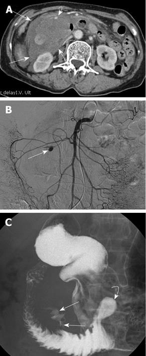
Figure 2 Diagnostic images on admission.
A: An approximately 7 cm sized acute duodenal diverticular hemorrhage (curved arrow) and surrounding large retroperitoneal hematoma (arrows) around the duodenum on contrast enhanced CT image; B: The causative arterial pseudoaneurysm (arrow) on superior mesenteric arteriogram; C: Barium leakage into the large hemorrhagic duodenal diverticulum (arrows) and another small silent diverticulum (curved arrow) in the duodenal third portion on upper gastrointestinal barium study.
- Citation: Kwon YJ, Kim JH, Kim SH, Kim BS, Kim HU, Choi EK, Jeong IH. Duodenal obstruction after successful embolization for duodenal diverticular hemorrhage: A case report. World J Gastroenterol 2009; 15(30): 3819-3822
- URL: https://www.wjgnet.com/1007-9327/full/v15/i30/3819.htm
- DOI: https://dx.doi.org/10.3748/wjg.15.3819