
















©2009 The WJG Press and Baishideng.
World J Gastroenterol. Jul 14, 2009; 15(26): 3312-3314
Published online Jul 14, 2009. doi: 10.3748/wjg.15.3312
Published online Jul 14, 2009. doi: 10.3748/wjg.15.3312
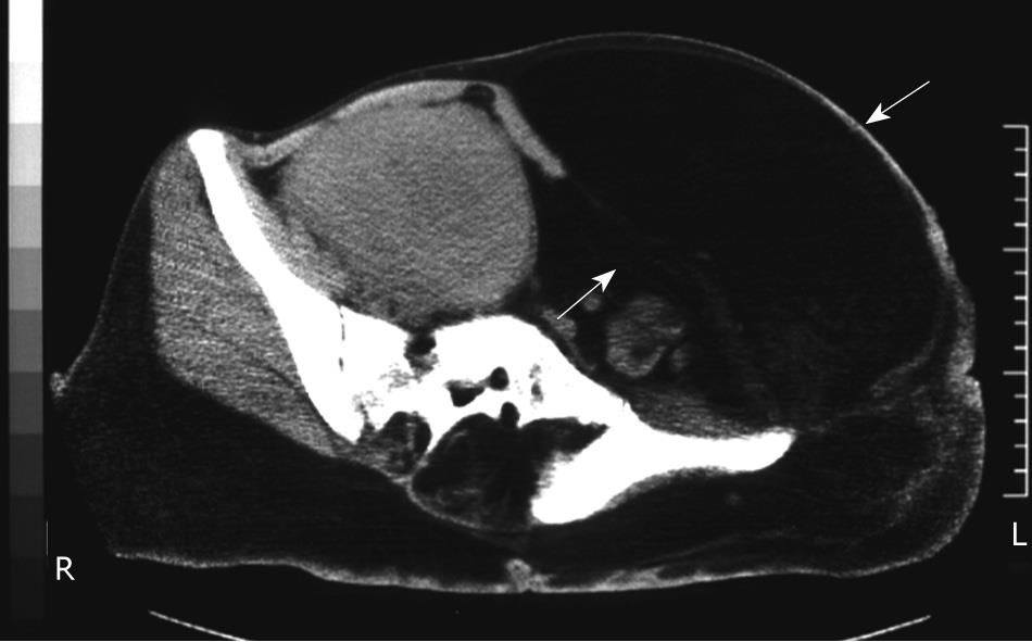
Figure 3 Preoperative computed tomography (CT) of the abdomen.
A large mass in the subcutaneous adipose tissue in the left lower abdominal wall was identified (arrows) and this encased the peritoneal organs to the right side.
- Citation: Nakayama Y, Kusuda S, Nagata N, Yamaguchi K. Excision of a large abdominal wall lipoma improved bowel passage in a Proteus syndrome patient. World J Gastroenterol 2009; 15(26): 3312-3314
- URL: https://www.wjgnet.com/1007-9327/full/v15/i26/3312.htm
- DOI: https://dx.doi.org/10.3748/wjg.15.3312