
















©2009 The WJG Press and Baishideng.
World J Gastroenterol. Apr 7, 2009; 15(13): 1656-1659
Published online Apr 7, 2009. doi: 10.3748/wjg.15.1656
Published online Apr 7, 2009. doi: 10.3748/wjg.15.1656
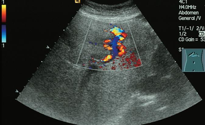
Figure 1 Color Doppler sonography of the portal vein aneurysm showing increased blood flow in the LPV and turbulence (arrow) at the distal portion of the aneurysmal sac.
- Citation: Lu ZY, Ao JY, Jiang TA, Peng ZY, Wang ZK. A large congenital and solitary intrahepatic arterioportal fistula in an old woman. World J Gastroenterol 2009; 15(13): 1656-1659
- URL: https://www.wjgnet.com/1007-9327/full/v15/i13/1656.htm
- DOI: https://dx.doi.org/10.3748/wjg.15.1656