
















©2008 The WJG Press and Baishideng.
World J Gastroenterol. Feb 21, 2008; 14(7): 1027-1033
Published online Feb 21, 2008. doi: 10.3748/wjg.14.1027
Published online Feb 21, 2008. doi: 10.3748/wjg.14.1027
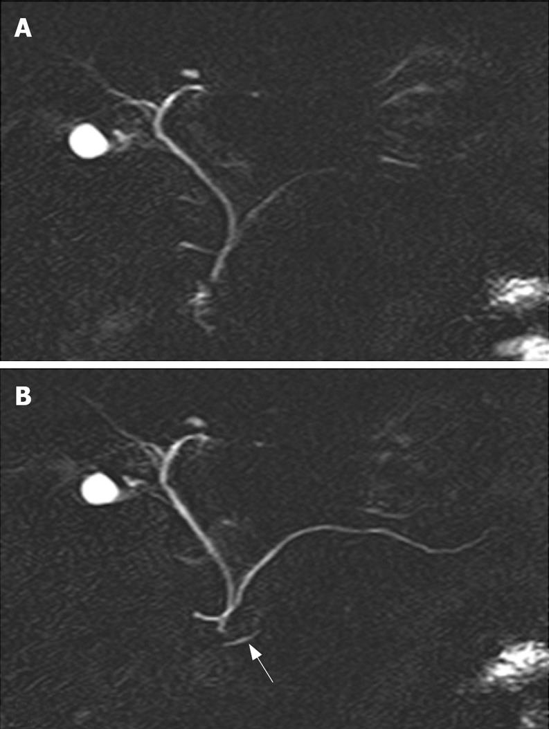
Figure 1 Normal pancreas divisum.
Unenhanced (A) and secretin-enhanced magnetic resonance cholangiopancreatography (B). The ventral pancreatic duct (arrow in B) and the entire course of the main dorsal pancreatic duct are seen only after secretin administration.
- Citation: Delhaye M, Matos C, Arvanitakis M, Devière J. Pancreatic ductal system obstruction and acute recurrent pancreatitis. World J Gastroenterol 2008; 14(7): 1027-1033
- URL: https://www.wjgnet.com/1007-9327/full/v14/i7/1027.htm
- DOI: https://dx.doi.org/10.3748/wjg.14.1027