
















©2008 The WJG Press and Baishideng.
World J Gastroenterol. Feb 14, 2008; 14(6): 944-947
Published online Feb 14, 2008. doi: 10.3748/wjg.14.944
Published online Feb 14, 2008. doi: 10.3748/wjg.14.944
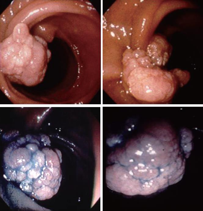
Figure 1 Upper gastrointestinal endoscopy in patient 1 revealed a semi-pedunculated polyp, 18 mm × 15 mm in diameter, on the oral side of the Papilla of Vater.
The polyp showed an irregularly lobular surface and slightly whitish color with diffusely scattered white spots on the surface.
- Citation: Suzuki S, Hirasaki S, Ikeda F, Yumoto E, Yamane H, Matsubara M. Three cases of solitary Peutz-Jeghers-type hamartomatous polyp in the duodenum. World J Gastroenterol 2008; 14(6): 944-947
- URL: https://www.wjgnet.com/1007-9327/full/v14/i6/944.htm
- DOI: https://dx.doi.org/10.3748/wjg.14.944