
















©2008 The WJG Press and Baishideng.
World J Gastroenterol. Feb 14, 2008; 14(6): 876-883
Published online Feb 14, 2008. doi: 10.3748/wjg.14.876
Published online Feb 14, 2008. doi: 10.3748/wjg.14.876
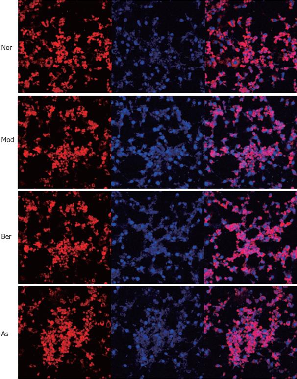
Figure 6 Fully differentiated cells were cultured for 12 h in serum-free DMEM with 0.
2% BSA and then they were, respectively, cultured for 24 h in DMEM containing 1% BSA for 24 h (Nor); or cultured for 24 h in DMEM containing 0.5 mmol/L PA and 1% BSA (Mod); or cultured for 48 h in DMEM containing 10 &mgr;mol/L Ber, 0.5 mmol/L PA, 1% BSA (Berberine); or cultured for 24h in DMEM containing 5 mmol/L aspirin, 0.5 mmol/L PA, 1% BSA (Aspirin). Without FFA stimulation NF-κB p65 is predominately found in the cytoplasm, imparting red color (Nor). While FFA stimulation, NF-κB p65 is translocated into the nucleus, showing pink color (Mod). The addition of 10 &mgr;mol/L Ber or 5 mmol/L aspirin clearly inhibits the FFAs induced NF-κB p65 translocation, as there is hardly any nuclear p65 staining found and in the figure, both red and pink colors can be observed (Berberine and Aspirin).
- Citation: Yi P, Lu FE, Xu LJ, Chen G, Dong H, Wang KF. Berberine reverses free-fatty-acid-induced insulin resistance in 3T3-L1 adipocytes through targeting IKKβ. World J Gastroenterol 2008; 14(6): 876-883
- URL: https://www.wjgnet.com/1007-9327/full/v14/i6/876.htm
- DOI: https://dx.doi.org/10.3748/wjg.14.876