
















©2008 The WJG Press and Baishideng.
World J Gastroenterol. Feb 14, 2008; 14(6): 853-863
Published online Feb 14, 2008. doi: 10.3748/wjg.14.853
Published online Feb 14, 2008. doi: 10.3748/wjg.14.853
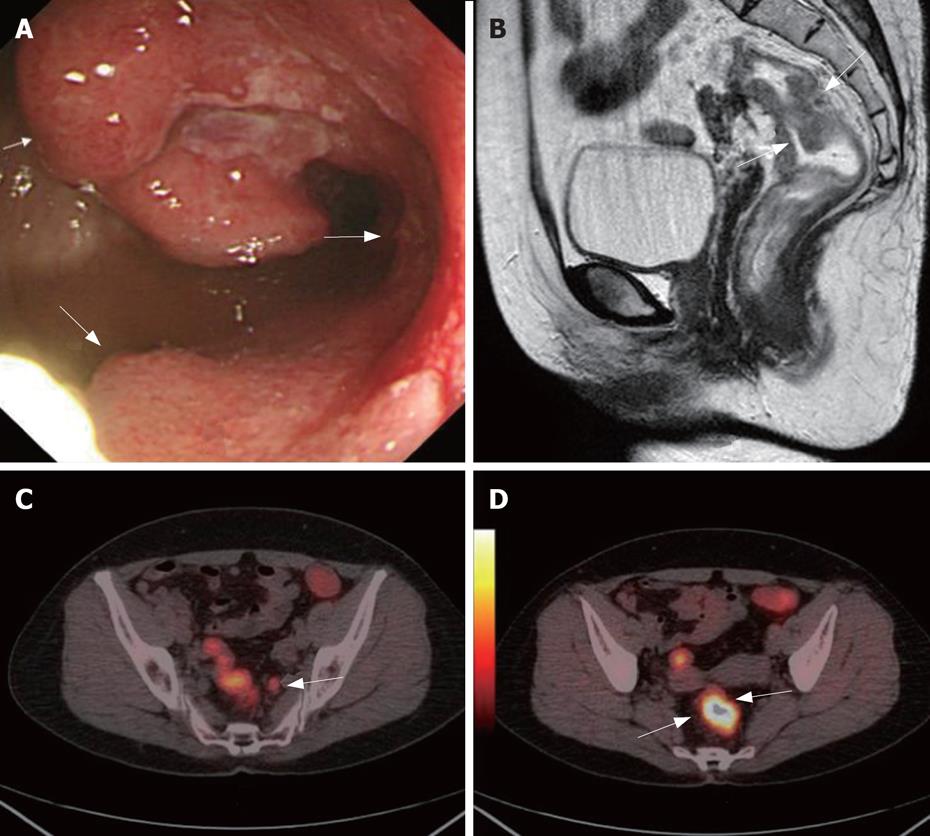
Figure 3 A 56-year-old women complained about blood stool for about 1 year.
CC showed one stenotic tumoral site in the rectum (white arrows, A). Sagittal MRC showed thickened rectum with suspected infiltration of the surrounding tissue (white arrows, B). Axial PET/CTC showed a high metabolism metastasis lymphoid node (white arrow, C). Axial PET/CTC revealed tumor sites with elevated glucose metabolism and clear circumscription (two white arrows, D). PET/CT also indicated no tumorous infiltration of the adjacent tissue and no distant metastases. This was later verified by histopathology. The case illustrated the value of VC in CRC TNM staging.
- Citation: Sun L, Wu H, Guan YS. Colonography by CT, MRI and PET/CT combined with conventional colonoscopy in colorectal cancer screening and staging. World J Gastroenterol 2008; 14(6): 853-863
- URL: https://www.wjgnet.com/1007-9327/full/v14/i6/853.htm
- DOI: https://dx.doi.org/10.3748/wjg.14.853