
















©2008 The WJG Press and Baishideng.
World J Gastroenterol. Dec 14, 2008; 14(46): 7127-7132
Published online Dec 14, 2008. doi: 10.3748/wjg.14.7127
Published online Dec 14, 2008. doi: 10.3748/wjg.14.7127
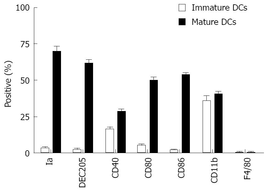
Figure 2 Immunophenotypic analysis of BM-DCs.
BM-DCs (2 × 10⁵-4 ×10⁵ cells) cultured for 5 d or 8 d were incubated with FITC- or PE-labeled MAbs. The phenotypes of these cells were analyzed by immunofluorescence staining as described in the Materials and Methods. The results are representative of three independent experiments and the data are given as mean ± SE.
-
Citation: Li YL, Wu YG, Wang YQ, Li Z, Wang RC, Wang L, Zhang YY. Bone marrow-derived dendritic cells pulsed with tumor lysates induce anti-tumor immunity against gastric cancer
ex vivo . World J Gastroenterol 2008; 14(46): 7127-7132 - URL: https://www.wjgnet.com/1007-9327/full/v14/i46/7127.htm
- DOI: https://dx.doi.org/10.3748/wjg.14.7127