
















©2008 The WJG Press and Baishideng.
World J Gastroenterol. Jul 7, 2008; 14(25): 4087-4090
Published online Jul 7, 2008. doi: 10.3748/wjg.14.4087
Published online Jul 7, 2008. doi: 10.3748/wjg.14.4087
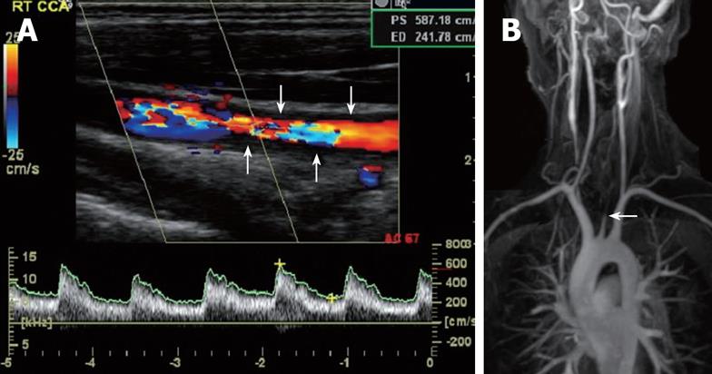
Figure 1 Ultrasound image of the right common carotid artery showing wall thickening (arrows) and blood flow with a significantly enhanced peak systolic velocity (PS > 587 cm/s), indicating a severe stenosis (A) and magnetic resonance angiography showing occlusion of the left common carotid (white arrows) and stenosis of the right common carotid (black arrows) arteries (B).
- Citation: Farrant MA, Mason JC, Wong NA, Longman RJ. Takayasu’s arteritis following Crohn’s disease in a young woman: Any evidence for a common pathogenesis? World J Gastroenterol 2008; 14(25): 4087-4090
- URL: https://www.wjgnet.com/1007-9327/full/v14/i25/4087.htm
- DOI: https://dx.doi.org/10.3748/wjg.14.4087