
















©2008 The WJG Press and Baishideng.
World J Gastroenterol. Jul 7, 2008; 14(25): 4005-4010
Published online Jul 7, 2008. doi: 10.3748/wjg.14.4005
Published online Jul 7, 2008. doi: 10.3748/wjg.14.4005
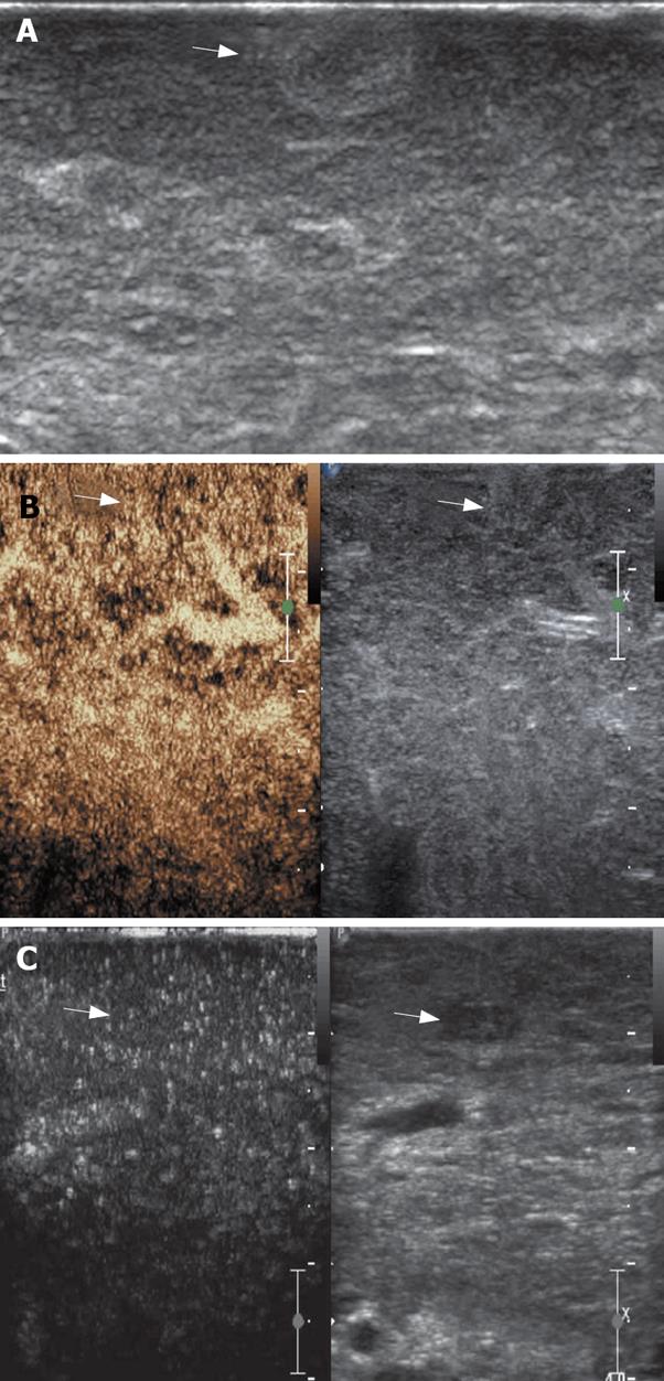
Figure 5 A: A hyperechoic nodule detected at IOUS in cirrhotic liver; B: The nodule shows no arterial enhancement and late parenchymal phase wash-out; C: A hypoechoic nodule detected at IOUS in cirrhotic liver, and it shows the same enhancing pattern with the surrounding parenchyma.
These two nodules were considered dysplastic and the diagnosis was confirmed by pathologic examination.
- Citation: Lu Q, Luo Y, Yuan CX, Zeng Y, Wu H, Lei Z, Zhong Y, Fan YT, Wang HH, Luo Y. Value of contrast-enhanced intraoperative ultrasound for cirrhotic patients with hepatocellular carcinoma: A report of 20 cases. World J Gastroenterol 2008; 14(25): 4005-4010
- URL: https://www.wjgnet.com/1007-9327/full/v14/i25/4005.htm
- DOI: https://dx.doi.org/10.3748/wjg.14.4005