
















©2008 The WJG Press and Baishideng.
World J Gastroenterol. May 14, 2008; 14(18): 2942-2945
Published online May 14, 2008. doi: 10.3748/wjg.14.2942
Published online May 14, 2008. doi: 10.3748/wjg.14.2942
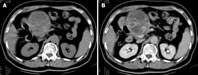
Figure 1 A: Pre-contrast CT shows a large, non-homogeneous, density mass in the head of the pancreas, with obvious atrophy in the body and tail of the pancreas; B: Lesion illustrates a well-defined, multilocular cystic mass with a cystic wall, fibrous septa and enhancement of solid components on post-contrast CT.
- Citation: Yu RS, Wang JW, Chen Y, Ding WH, Xu XF, Chen LR. A case of primary malignant fibrous histiocytoma of the pancreas: CT and MRI findings. World J Gastroenterol 2008; 14(18): 2942-2945
- URL: https://www.wjgnet.com/1007-9327/full/v14/i18/2942.htm
- DOI: https://dx.doi.org/10.3748/wjg.14.2942