
















©2008 The WJG Press and Baishideng.
World J Gastroenterol. May 14, 2008; 14(18): 2912-2914
Published online May 14, 2008. doi: 10.3748/wjg.14.2912
Published online May 14, 2008. doi: 10.3748/wjg.14.2912
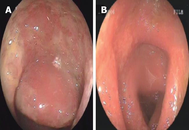
Figure 1 Colonoscopic appearance of the mucosa before (A) and during the 14 d of anti-viral treatment (B).
The presence of edema and redness is seen in both images. Note the ulcers in the sigmoid colon before the treatment as shown in A.
- Citation: Sari I, Birlik M, Gonen C, Akar S, Gurel D, Onen F, Akkoc N. Cytomegalovirus colitis in a patient with Behcet’s disease receiving tumor necrosis factor alpha inhibitory treatment. World J Gastroenterol 2008; 14(18): 2912-2914
- URL: https://www.wjgnet.com/1007-9327/full/v14/i18/2912.htm
- DOI: https://dx.doi.org/10.3748/wjg.14.2912