
















©2008 The WJG Press and Baishideng.
World J Gastroenterol. Apr 28, 2008; 14(16): 2602-2608
Published online Apr 28, 2008. doi: 10.3748/wjg.14.2602
Published online Apr 28, 2008. doi: 10.3748/wjg.14.2602
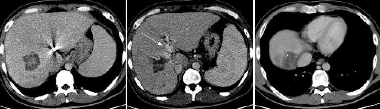
Figure 2 After three courses of GEMFP, CT scanning showed marked regression of the primary tumor (55 mm) with demonstrable portal blood flow (arrow) and the disappearance of hepatic venous tumor thrombus in the inferior vena cava.
- Citation: Uka K, Aikata H, Takaki S, Kawaoka T, Saneto H, Miki D, Takahashi S, Toyota N, Ito K, Chayama K. Systemic gemcitabine combined with intra-arterial low-dose cisplatin and 5-fluorouracil for advanced hepatocellular carcinoma: Seven cases. World J Gastroenterol 2008; 14(16): 2602-2608
- URL: https://www.wjgnet.com/1007-9327/full/v14/i16/2602.htm
- DOI: https://dx.doi.org/10.3748/wjg.14.2602