
















©2007 Baishideng Publishing Group Co.
World J Gastroenterol. Jan 21, 2007; 13(3): 448-451
Published online Jan 21, 2007. doi: 10.3748/wjg.v13.i3.448
Published online Jan 21, 2007. doi: 10.3748/wjg.v13.i3.448
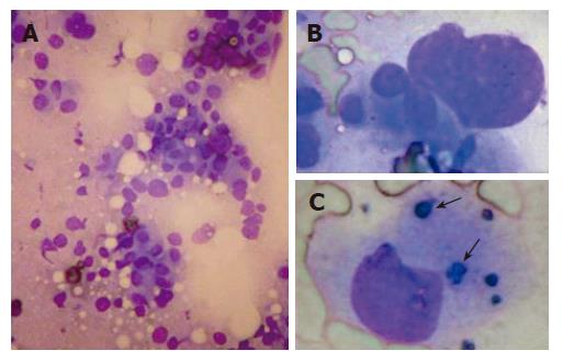
Figure 2 Cytodiagnosis of hepatocellular carcinoma by quick Liu stain showing scattered distribution of malignant cells bearing atypical naked (A) and large nuclei (B) with scanty cytoplasm but without trabecular structure and sinusoids (A: 100 x, B: 400 x) and bile pigmentation (arrows) in cytoplasma (C) (400 x).
- Citation: Changchien CS, Wang JH, Lu SN, Hung CH, Chen CH, Lee CM. Liu-stain quick cytodiagnosis of ultrasound-guided fine needle aspiration in diagnosis of liver tumors. World J Gastroenterol 2007; 13(3): 448-451
- URL: https://www.wjgnet.com/1007-9327/full/v13/i3/448.htm
- DOI: https://dx.doi.org/10.3748/wjg.v13.i3.448