
















©2007 Baishideng Publishing Group Co.
World J Gastroenterol. Jun 14, 2007; 13(22): 3141-3143
Published online Jun 14, 2007. doi: 10.3748/wjg.v13.i22.3141
Published online Jun 14, 2007. doi: 10.3748/wjg.v13.i22.3141
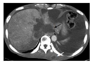
Figure 1 Computed tomo-graphy shows a large mass with rim enhancement around intrahepatic portion IVC of liver which encircles IVC (black arrow).
Metastatic lesions seen in the left spinal erector muscle (white arrow) and right psoas muscle (not shown).
- Citation: Kwon OS, Jun DW, Kim SH, Chung MY, Kim NI, Song MH, Lee HH, Kim SH, Jo YJ, Park YS, Joo JE. Distant skeletal muscle metastasis from intrahepatic cholangiocarcinoma presenting as Budd-Chiari syndrome. World J Gastroenterol 2007; 13(22): 3141-3143
- URL: https://www.wjgnet.com/1007-9327/full/v13/i22/3141.htm
- DOI: https://dx.doi.org/10.3748/wjg.v13.i22.3141