
















©2007 Baishideng Publishing Group Co.
World J Gastroenterol. Jun 14, 2007; 13(22): 3112-3116
Published online Jun 14, 2007. doi: 10.3748/wjg.v13.i22.3112
Published online Jun 14, 2007. doi: 10.3748/wjg.v13.i22.3112
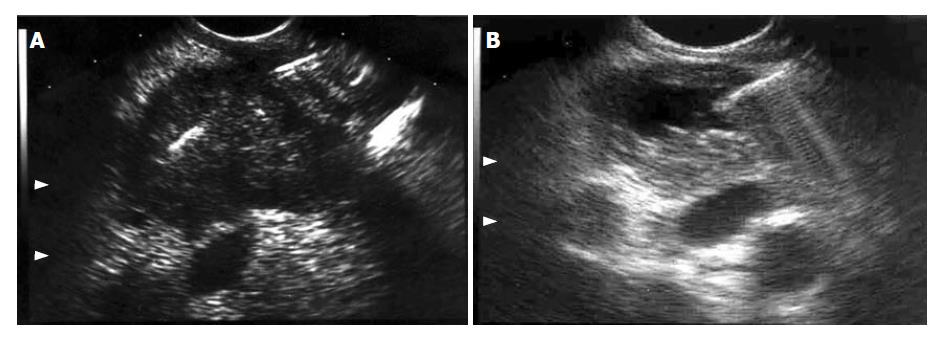
Figure 1 EUS images during FNA of (A) solid pancreatic tumor (colon carcinoma metastasis), and (B) cystic lesion (cystadenocarcinoma), both confirmed by the cytopathological assessment.
Note the needle inside both lesions.
- Citation: Ardengh JC, Lopes CV, de Lima LFP, de Oliveira JR, Venco F, Santo GC, Módena JLP. Diagnosis of pancreatic tumors by endoscopic ultrasound-guided fine-needle aspiration. World J Gastroenterol 2007; 13(22): 3112-3116
- URL: https://www.wjgnet.com/1007-9327/full/v13/i22/3112.htm
- DOI: https://dx.doi.org/10.3748/wjg.v13.i22.3112