
















©2007 Baishideng Publishing Group Co.
World J Gastroenterol. May 21, 2007; 13(19): 2747-2751
Published online May 21, 2007. doi: 10.3748/wjg.v13.i19.2747
Published online May 21, 2007. doi: 10.3748/wjg.v13.i19.2747
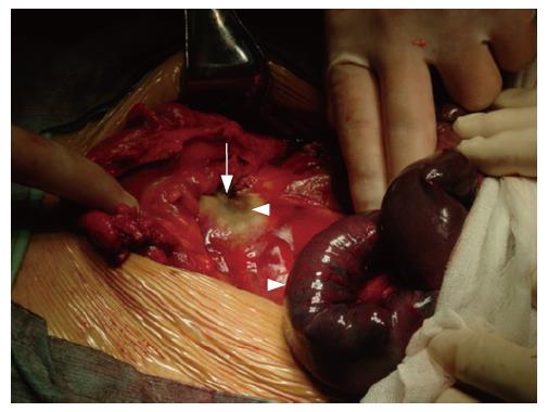
Figure 3 The target tumor presented as a white coagulative necrosis in the exploratory laparotomy 3 d after HIFU (arrowhead), the purple ischemia of the necrotic small intestines are also shown (arrowhead), and the SMA turned out from the center of tumor (arrow).
- Citation: Li JJ, Xu GL, Gu MF, Luo GY, Rong Z, Wu PH, Xia JC. Complications of high intensity focused ultrasound in patients with recurrent and metastatic abdominal tumors. World J Gastroenterol 2007; 13(19): 2747-2751
- URL: https://www.wjgnet.com/1007-9327/full/v13/i19/2747.htm
- DOI: https://dx.doi.org/10.3748/wjg.v13.i19.2747