
















©2007 Baishideng Publishing Group Co.
World J Gastroenterol. Mar 14, 2007; 13(10): 1632-1635
Published online Mar 14, 2007. doi: 10.3748/wjg.v13.i10.1632
Published online Mar 14, 2007. doi: 10.3748/wjg.v13.i10.1632
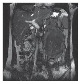
Figure 2 Coronal MRI image demonstrating a tumor with low to intermediate mixed signal intensity in the lesion's periphery and low signal intensity in the central region of the lesion on T2 weighted images.
- Citation: Sun L, Wu H, Zhuang YZ, Guan YS. A rare case of pregnancy complicated by mesenteric mass: What does chylous ascites tell us? World J Gastroenterol 2007; 13(10): 1632-1635
- URL: https://www.wjgnet.com/1007-9327/full/v13/i10/1632.htm
- DOI: https://dx.doi.org/10.3748/wjg.v13.i10.1632