
















©2006 Baishideng Publishing Group Co.
World J Gastroenterol. Oct 28, 2006; 12(40): 6556-6558
Published online Oct 28, 2006. doi: 10.3748/wjg.v12.i40.6556
Published online Oct 28, 2006. doi: 10.3748/wjg.v12.i40.6556
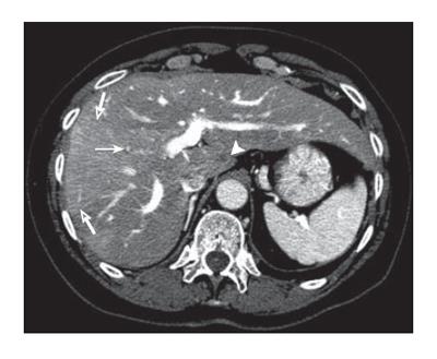
Figure 2 Contrast enhanced CT at the level of the portal bifurcation after treatment shows improved hepatic metastasis in segment I (arrowhead), non-visualization of the thrombosed anterior right segmental portal vein (arrow), opacified posterior segmental portal veins, middle and right hepatic veins, and wedge shaped increased enhancement (open arrows) of the anterior sector secondary to portal vein occlusion.
- Citation: Donadon M, Vauthey JN, Loyer EM, Charnsangavej C, Abdalla EK. Portal thrombosis and steatosis after preoperativechemotherapy with FOLFIRI-bevacizumab for colorectal liver metastases. World J Gastroenterol 2006; 12(40): 6556-6558
- URL: https://www.wjgnet.com/1007-9327/full/v12/i40/6556.htm
- DOI: https://dx.doi.org/10.3748/wjg.v12.i40.6556