
















©2006 Baishideng Publishing Group Co.
World J Gastroenterol. Oct 28, 2006; 12(40): 6473-6499
Published online Oct 28, 2006. doi: 10.3748/wjg.v12.i40.6473
Published online Oct 28, 2006. doi: 10.3748/wjg.v12.i40.6473
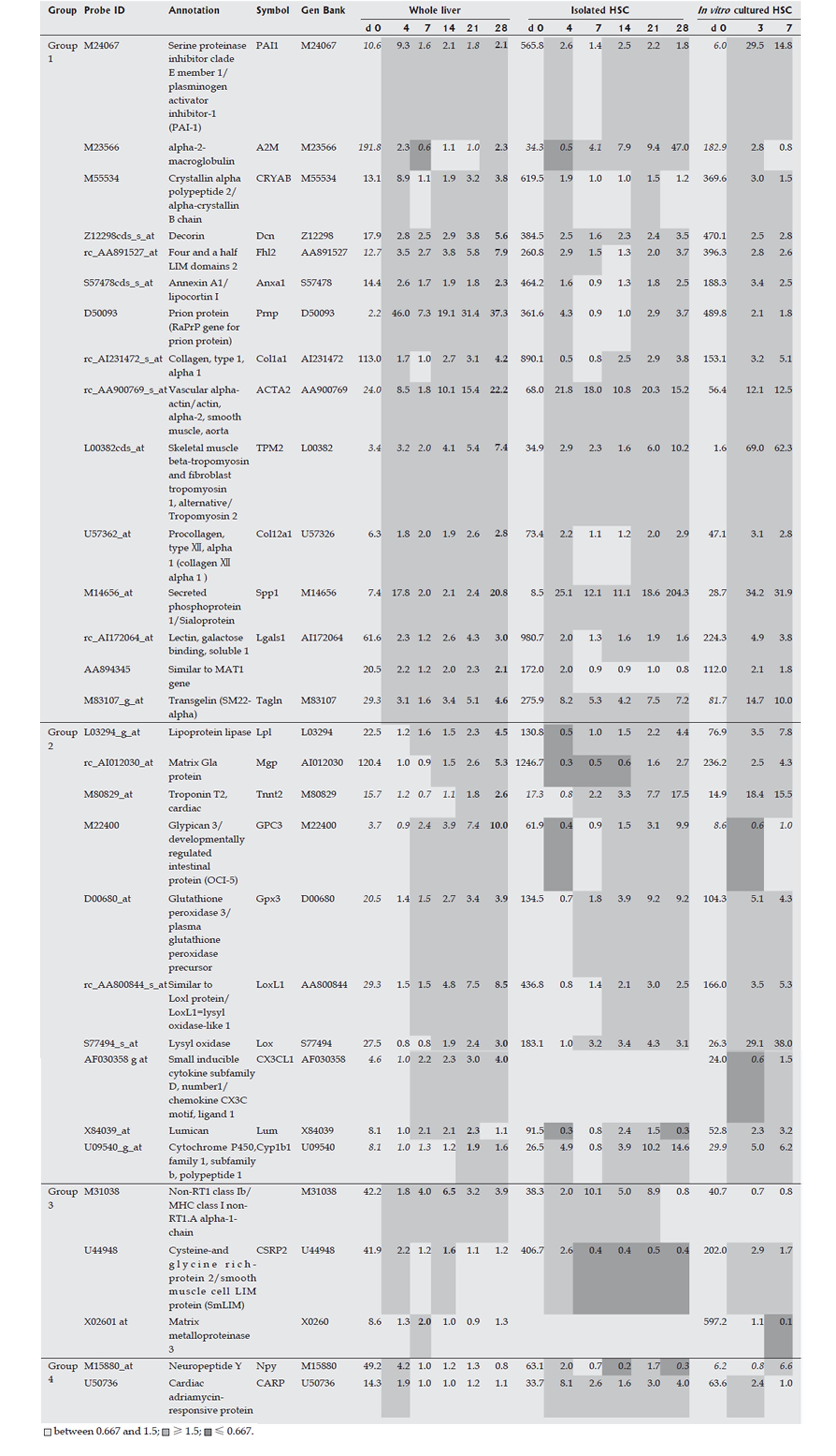
Figure 3 Marker genes in hepatic stellate cells (HSC).
Expression profiles of the whole liver, of isolated HSCs during fibrogenesis, and of in vitro cultured isolated HSCs were obtained using a rat Genome U34A Array (Affymetrix). Marker genes for HSCs are listed. Expression intensities are shown for d 0, and expression intensity data for d 4, 7, 14, 21 and 28 are displayed as ratios to the d 0 expression levels for the analysis of whole liver and isolated HSCs. For in vitro cultured HSCs, the d 0 data are similarly shown as expression intensities, and data on d 4 and 7 are shown as ratios to the expression levels on d 0. Italics indicate an “absent” call by the Affymetrix software, and bold text indicates the highest ratio in the chronic phase (d 7, 14, 21 and 28).
- Citation: Takahara Y, Takahashi M, Wagatsuma H, Yokoya F, Zhang QW, Yamaguchi M, Aburatani H, Kawada N. Gene expression profiles of hepatic cell-type specific marker genes in progression of liver fibrosis. World J Gastroenterol 2006; 12(40): 6473-6499
- URL: https://www.wjgnet.com/1007-9327/full/v12/i40/6473.htm
- DOI: https://dx.doi.org/10.3748/wjg.v12.i40.6473