
















©2006 Baishideng Publishing Group Co.
World J Gastroenterol. Oct 21, 2006; 12(39): 6405-6407
Published online Oct 21, 2006. doi: 10.3748/wjg.v12.i39.6405
Published online Oct 21, 2006. doi: 10.3748/wjg.v12.i39.6405
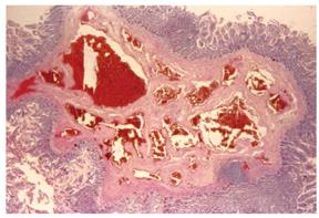
Figure 3 Histological section showing enteric wall mucosa and angiomatous lesions through the muscular layer of the colonic wall (HE x 25).
- Citation: Marinis A, Kairi E, Theodosopoulos T, Kondi-Pafiti A, Smyrniotis V. Right colon and liver hemangiomatosis: A case report and a review of the literature. World J Gastroenterol 2006; 12(39): 6405-6407
- URL: https://www.wjgnet.com/1007-9327/full/v12/i39/6405.htm
- DOI: https://dx.doi.org/10.3748/wjg.v12.i39.6405