
















©2006 Baishideng Publishing Group Co.
World J Gastroenterol. Mar 28, 2006; 12(12): 1933-1936
Published online Mar 28, 2006. doi: 10.3748/wjg.v12.i12.1933
Published online Mar 28, 2006. doi: 10.3748/wjg.v12.i12.1933
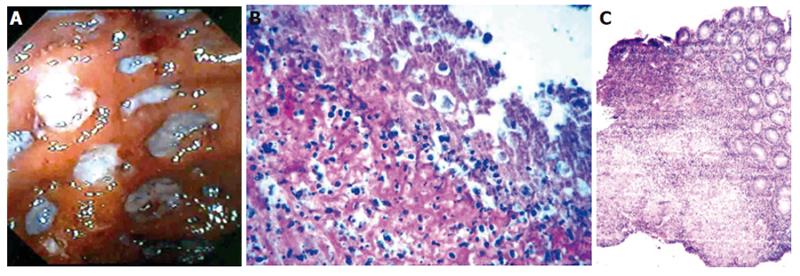
Figure 4 Colonoscopy findings in a patient showing deep round and oval ulcers in the cecum (A), histological examination of the biopsies obtained from ulcers in the cecum showing the presence of trophozoites of E.
histolytica (B) and from a patient with amebic liver abscess and cecal mass showing the presence of multiple epithelioid cell granulomas and Langhans giant cells (C) (H & E X 40).
- Citation: Misra SP, Misra V, Dwivedi M. Ileocecal masses in patients with amebic liver abscess: Etiology and management. World J Gastroenterol 2006; 12(12): 1933-1936
- URL: https://www.wjgnet.com/1007-9327/full/v12/i12/1933.htm
- DOI: https://dx.doi.org/10.3748/wjg.v12.i12.1933