
















©2005 Baishideng Publishing Group Inc.
World J Gastroenterol. Dec 21, 2005; 11(47): 7444-7449
Published online Dec 21, 2005. doi: 10.3748/wjg.v11.i47.7444
Published online Dec 21, 2005. doi: 10.3748/wjg.v11.i47.7444
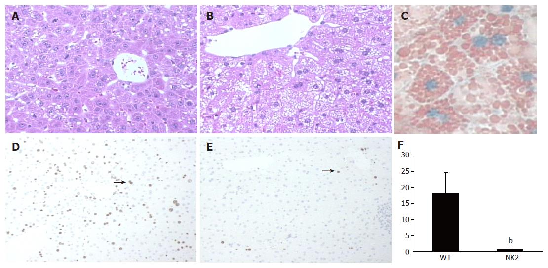
Figure 2 Liver regeneration and proliferation of hepatocytes after PH.
H-E stain of WT mouse liver (A) and NK2 transgenic mouse liver (B) at 48 h after PH. NK2 transgenic mouse liver contained a large amount of intracellular small droplets. Magnification, ×200. Oil-red-O stain of NK2 transgenic mouse liver (C) at 48 h after PH. Note the presence of many small lipid droplets in hepatocytes. Magnification, ×400. BrdU stain of WT mouse liver (D) and NK2 transgenic mouse liver (E) at 48 h after PH. The arrows show hepatocytes undergoing DNA synthesis. Magnification, ×100. The BrdU labeling index of WT mice and NK2 transgenic mice at 48 h after PH (n = 30 per group). (F) Data are mean±SD of the mean. Hepatocyte proliferation was significantly reduced in NK2 transgenic mouse liver (bP<0.01 vs WT mouse liver).
- Citation: Otsuka T, Horiguchi N, Kanda D, Kosone T, Yamazaki Y, Yuasa K, Sohara N, Kakizaki S, Sato K, Takagi H, Merlino G, Mori M. Overexpression of NK2 inhibits liver regeneration after partial hepatectomy in mice. World J Gastroenterol 2005; 11(47): 7444-7449
- URL: https://www.wjgnet.com/1007-9327/full/v11/i47/7444.htm
- DOI: https://dx.doi.org/10.3748/wjg.v11.i47.7444