
















©The Author(s) 2005.
World J Gastroenterol. Oct 21, 2005; 11(39): 6225-6227
Published online Oct 21, 2005. doi: 10.3748/wjg.v11.i39.6225
Published online Oct 21, 2005. doi: 10.3748/wjg.v11.i39.6225
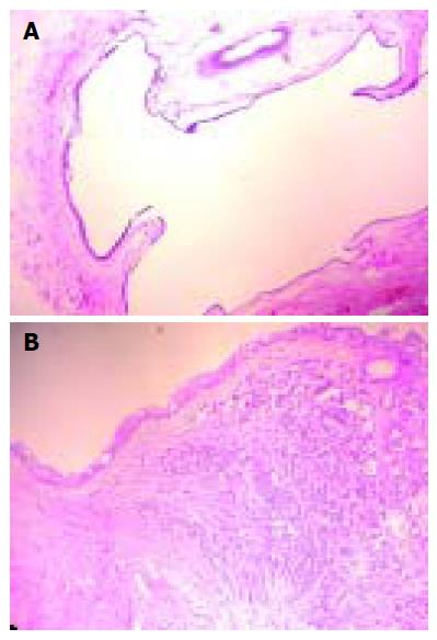
Figure 3 A and B Showing malignant alteration of the cyst histologically presenting adenosquamous carcinoma, mostly well differentiated adenocarcinoma admixed with foci of squamous differentiation or poorly differentiated areas and clear resection margins.
- Citation: Krivokapic Z, Dimitrijevic I, Barisic G, Markovic V, Krstic M. Adenosquamous carcinoma arising within a retrorectal tailgut cyst: Report of a case. World J Gastroenterol 2005; 11(39): 6225-6227
- URL: https://www.wjgnet.com/1007-9327/full/v11/i39/6225.htm
- DOI: https://dx.doi.org/10.3748/wjg.v11.i39.6225