
















©The Author(s) 2005.
World J Gastroenterol. Sep 28, 2005; 11(36): 5742-5745
Published online Sep 28, 2005. doi: 10.3748/wjg.v11.i36.5742
Published online Sep 28, 2005. doi: 10.3748/wjg.v11.i36.5742
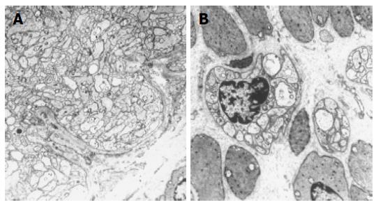
Figure 6 Electron microscopic sections show thickened parasympathetic nerve fibers in the distal part of the resected segment (A) and normal numbers of ganglia in the proximal part (B).
- Citation: Werner CR, Stoltenburg-Didinger G, Weidemann H, Benckert C, Schmidtmann M, Voort IRVD, Andresen V, Klapp BF, Neuhaus P, Wiedenmann B, Mönnikes H. Megacolon in adulthood after surgical treatment of Hirschsprung’s disease in early childhood. World J Gastroenterol 2005; 11(36): 5742-5745
- URL: https://www.wjgnet.com/1007-9327/full/v11/i36/5742.htm
- DOI: https://dx.doi.org/10.3748/wjg.v11.i36.5742