
















©2005 Baishideng Publishing Group Inc.
World J Gastroenterol. Sep 21, 2005; 11(35): 5577-5581
Published online Sep 21, 2005. doi: 10.3748/wjg.v11.i35.5577
Published online Sep 21, 2005. doi: 10.3748/wjg.v11.i35.5577
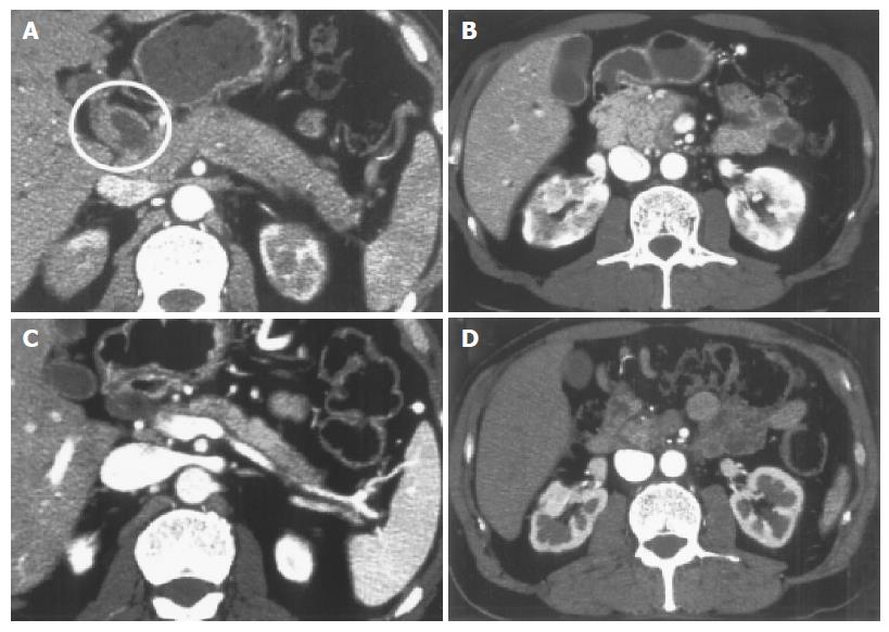
Figure 1 Abdominal CT scans on admission.
Note the diffuse enlargement of the pancreas and wall thickness of the enlarged common bile duct (CBD) (white circle) (A, B). Abdominal CT scans taken at 3 mo after treatment. Note the improvement in pancreatic swelling and CBD (C, D).
- Citation: Taguchi M, Aridome G, Abe S, Kume K, Tashiro M, Yamamoto M, Kihara Y, Nakamura H, Otsuki M. Autoimmune pancreatitis with IgG4-positive plasma cell infiltration in salivary glands and biliary tract. World J Gastroenterol 2005; 11(35): 5577-5581
- URL: https://www.wjgnet.com/1007-9327/full/v11/i35/5577.htm
- DOI: https://dx.doi.org/10.3748/wjg.v11.i35.5577