
















©The Author(s) 2005.
World J Gastroenterol. Jul 21, 2005; 11(27): 4281-4284
Published online Jul 21, 2005. doi: 10.3748/wjg.v11.i27.4281
Published online Jul 21, 2005. doi: 10.3748/wjg.v11.i27.4281
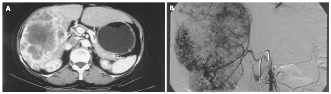
Figure 1 A: Liver CT scan showed a huge tumor in the right lobe of the liver with enhancement from the periphery in the arterial phase; B: The hepatic arteriogram demonstrated numerous feeding vessels surrounding and entering into the tumor.
The patient had variation of the right hepatic artery from the superior mesenteric artery.
- Citation: Kim BW, Wang HJ, Jeong IH, Ahn SI, Kim MW. Metastatic liver cancer: A rare case. World J Gastroenterol 2005; 11(27): 4281-4284
- URL: https://www.wjgnet.com/1007-9327/full/v11/i27/4281.htm
- DOI: https://dx.doi.org/10.3748/wjg.v11.i27.4281