
















©The Author(s) 2005.
World J Gastroenterol. Jul 14, 2005; 11(26): 4114-4116
Published online Jul 14, 2005. doi: 10.3748/wjg.v11.i26.4114
Published online Jul 14, 2005. doi: 10.3748/wjg.v11.i26.4114
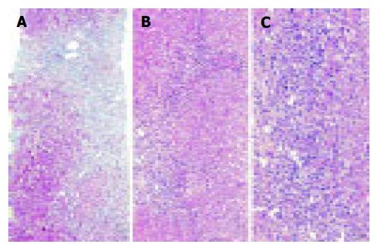
Figure 2 A liver biopsy obtained 3 mo after the first clinical presentation of the patient showed marked bridging fibrosis (A).
A moderate chronic inflammatory infiltrate with few scattered eosinophils was found in the septa (B), and an interface hepatitis was also noted (C). The histopathological findings were compatible with AIH. Masson trichrome stain (A) and H&E-stain (B and C). Original magnifications 100x (A and B) and 200x (C).
- Citation: Csepregi A, Treiber G, Röcken C, Malfertheiner P. Acute exacerbation of autoimmune hepatitis induced by Twinrix. World J Gastroenterol 2005; 11(26): 4114-4116
- URL: https://www.wjgnet.com/1007-9327/full/v11/i26/4114.htm
- DOI: https://dx.doi.org/10.3748/wjg.v11.i26.4114