
















©2005 Baishideng Publishing Group Inc.
World J Gastroenterol. Jun 28, 2005; 11(24): 3797-3799
Published online Jun 28, 2005. doi: 10.3748/wjg.v11.i24.3797
Published online Jun 28, 2005. doi: 10.3748/wjg.v11.i24.3797
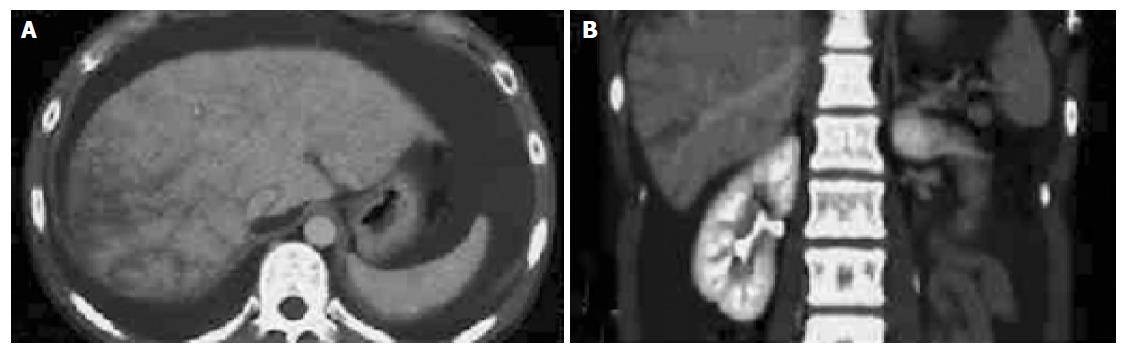
Figure 1 Computerized tomography scan of the abdomen.
(A) and (B) show the sagittal and coronal images of abdomen, respectively. Hepatomegaly with inhomogeneous distribution of contrast medium is visualized. Massive ascites is also recognized.
- Citation: Araki Y, Sakaguchi C, Ishizuka I, Sasaki M, Tsujikawa T, Koyama S, Furukawa A, Fujiyama Y. Budd-Chiari syndrome: A case with a combination of hepatic vein and superior vena cava occlusion. World J Gastroenterol 2005; 11(24): 3797-3799
- URL: https://www.wjgnet.com/1007-9327/full/v11/i24/3797.htm
- DOI: https://dx.doi.org/10.3748/wjg.v11.i24.3797