
















©2005 Baishideng Publishing Group Inc.
World J Gastroenterol. Jun 28, 2005; 11(24): 3710-3713
Published online Jun 28, 2005. doi: 10.3748/wjg.v11.i24.3710
Published online Jun 28, 2005. doi: 10.3748/wjg.v11.i24.3710
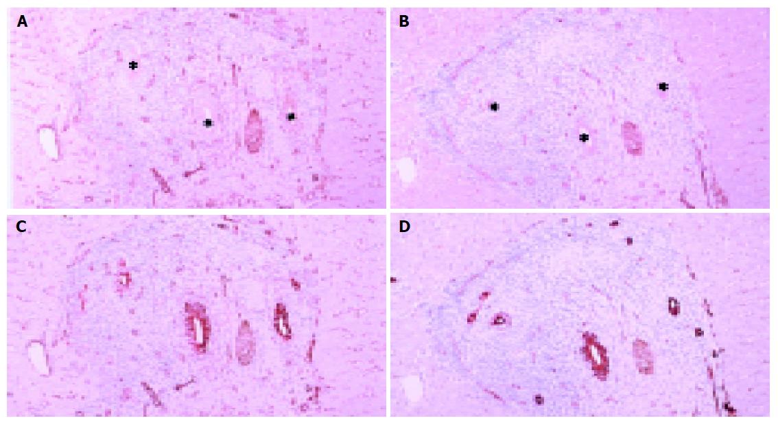
Figure 2 Light microscopic distributions of immunohistochemically stained CAV-1 and -2 in PBC stage 1 liver tissue.
A: CAV-1, ×100; B: CAV-2, ×100; C: double staining of CAV-1 and cytokeratin 7, ×100; D: double staining of CAV-2 and cytokeratin 7, ×100. Hematoxylin counterstain. Immunostaining of CAV-1 and -2 is observed to some degree on interlobular bile ducts, intense immunostaining on proliferative bile ductules, and some immunostaining on hepatic sinusoidal lining cells. Asterisks denote interlobular bile ducts.
- Citation: Yokomori H, Oda M, Wakabayashi G, Kitajima M, Yoshimura K, Nomura M, Hibi T. High expressions of caveolins on the proliferating bile ductules in primary biliary cirrhosis. World J Gastroenterol 2005; 11(24): 3710-3713
- URL: https://www.wjgnet.com/1007-9327/full/v11/i24/3710.htm
- DOI: https://dx.doi.org/10.3748/wjg.v11.i24.3710