
















©2005 Baishideng Publishing Group Inc.
World J Gastroenterol. May 7, 2005; 11(17): 2603-2608
Published online May 7, 2005. doi: 10.3748/wjg.v11.i17.2603
Published online May 7, 2005. doi: 10.3748/wjg.v11.i17.2603
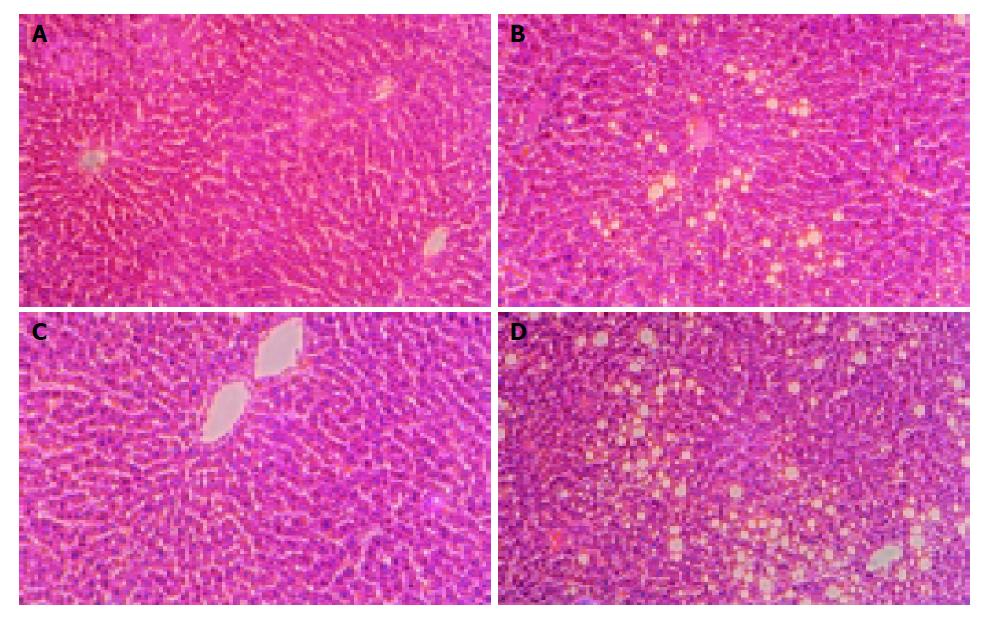
Figure 2 Histological examinations of specimens from the liver in each group.
Livers from group MC (A), ME (B), FC (C), and FE (D) are shown. Sections of the liver from rats fed with control diet for 12 wk show normal histology with no evidence of pathologic change (A and C). Fat accumulation in female ethanol-fed rats was panlobular (D), but it was seen in pericentral areas in male ethanol-fed rats (B). (H&E stain, magnification: ×200).
- Citation: Huang CC, Chen JR, Liu CC, Chen KT, Shieh MJ, Yang SC. Effects of long-term ethanol consumption on jejunal lipase and disaccharidase activities in male and female rats. World J Gastroenterol 2005; 11(17): 2603-2608
- URL: https://www.wjgnet.com/1007-9327/full/v11/i17/2603.htm
- DOI: https://dx.doi.org/10.3748/wjg.v11.i17.2603