
















©2005 Baishideng Publishing Group Inc.
World J Gastroenterol. Apr 21, 2005; 11(15): 2269-2276
Published online Apr 21, 2005. doi: 10.3748/wjg.v11.i15.2269
Published online Apr 21, 2005. doi: 10.3748/wjg.v11.i15.2269
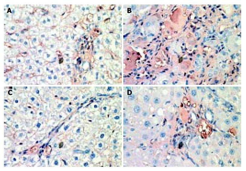
Figure 3 Immunohistochemical staining for TGF-β1 in liver sections.
×400 for A, B, C and D. (A) TGF-β1 immunoreactivity was evenly distributed within the interstitial tissue and liver sinusoidal in the normal group; (B) Stout connective tissue septa were strongly immunoreactive for TGF-β1 in the model group; (C) The staining intensity was much weaker in JinSanE group I than that in the model group; and (D) The staining positivity in JinSanE group II was stronger than that in the JinSanE group II, but still less than that in the model group.
-
Citation: Song SL, Gong ZJ, Zhang QR, Huang TX. Effects of Chinese traditional compound, JinSanE, on expression of TGF-β1 and TGF-β1 type II receptor mRNA, Smad3 and Smad7 on experimental hepatic fibrosis
in vivo . World J Gastroenterol 2005; 11(15): 2269-2276 - URL: https://www.wjgnet.com/1007-9327/full/v11/i15/2269.htm
- DOI: https://dx.doi.org/10.3748/wjg.v11.i15.2269