
















©The Author(s) 2004.
World J Gastroenterol. Mar 15, 2004; 10(6): 864-870
Published online Mar 15, 2004. doi: 10.3748/wjg.v10.i6.864
Published online Mar 15, 2004. doi: 10.3748/wjg.v10.i6.864
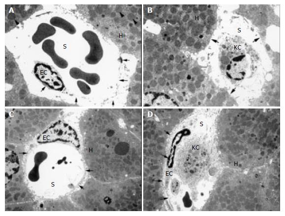
Figure 2 Transmission electron microphotographs of untreated and GSH- treated liver allografts.
Arrow-heads indicate mito-chondrial swelling of hepatocytes in untreated livers (A) which was almost absent in allografts treated with 100 µmol GSH/(h·kg) during reperfusion (C). Arrows demonstrate the loss of hepatocyte microvilli in untreated controls (B) whereas GSH administra-tion preserved microvilli in nearly all hepatocyte membranes (D). As indicated by open arrows detachment of SEC (A) as well as its complete loss (B) was evident in untreated livers. GSH- treatment preserved sinusoidal endothelial lining with a normal space of Dissé (C,D). Letters indicate: H, hepatocyte; KC, Kupffer cell; EC, endothelial cell; S, sinusoidal lumen. Bars represent either 1.7 µm (B) or 2.5 µm (A,C,D).
- Citation: Schauer RJ, Kalmuk S, Gerbes AL, Leiderer R, Meissner H, Schildberg FW, Messmer K, Bilzer M. Intravenous administration of glutathione protects parenchymal and non-parenchymal liver cells against reperfusion injury following rat liver transplantation. World J Gastroenterol 2004; 10(6): 864-870
- URL: https://www.wjgnet.com/1007-9327/full/v10/i6/864.htm
- DOI: https://dx.doi.org/10.3748/wjg.v10.i6.864