
















©The Author(s) 2004.
World J Gastroenterol. Sep 15, 2004; 10(18): 2767-2768
Published online Sep 15, 2004. doi: 10.3748/wjg.v10.i18.2767
Published online Sep 15, 2004. doi: 10.3748/wjg.v10.i18.2767
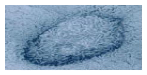
Figure 3 Scanning electron microscopy of the Peyer’s patches demonstrating M cells with microfolds.
- Citation: Ishimoto H, Isomoto H, Shikuwa S, Wen CY, Suematu T, Ito M, Murata I, Ishibashi H, Kohno S. Endoscopic identification of Peyer’s patches of the terminal ileum in a patient with Crohn’s disease. World J Gastroenterol 2004; 10(18): 2767-2768
- URL: https://www.wjgnet.com/1007-9327/full/v10/i18/2767.htm
- DOI: https://dx.doi.org/10.3748/wjg.v10.i18.2767