Copyright
©The Author(s) 2025.
Artif Intell Gastrointest Endosc. Jun 8, 2025; 6(2): 106600
Published online Jun 8, 2025. doi: 10.37126/aige.v6.i2.106600
Published online Jun 8, 2025. doi: 10.37126/aige.v6.i2.106600
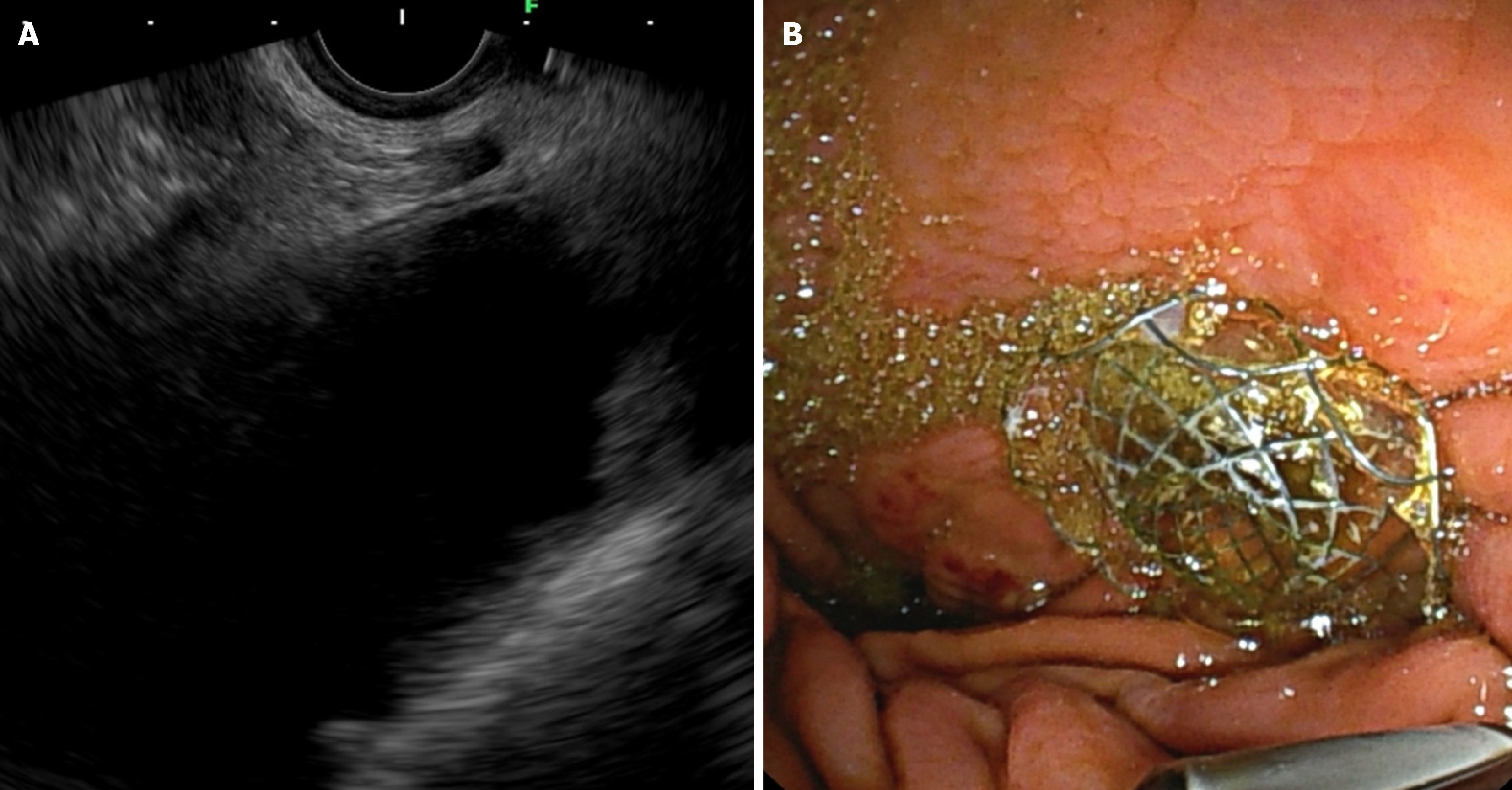
Figure 1 Endoscopic ultrasound-guided gastroenterostomy in afferent loop syndrome.
А: Echoendoscopic view demonstrating the afferent loop; В: Fluoroscopic view confirming the positioning and deployment of the stent.
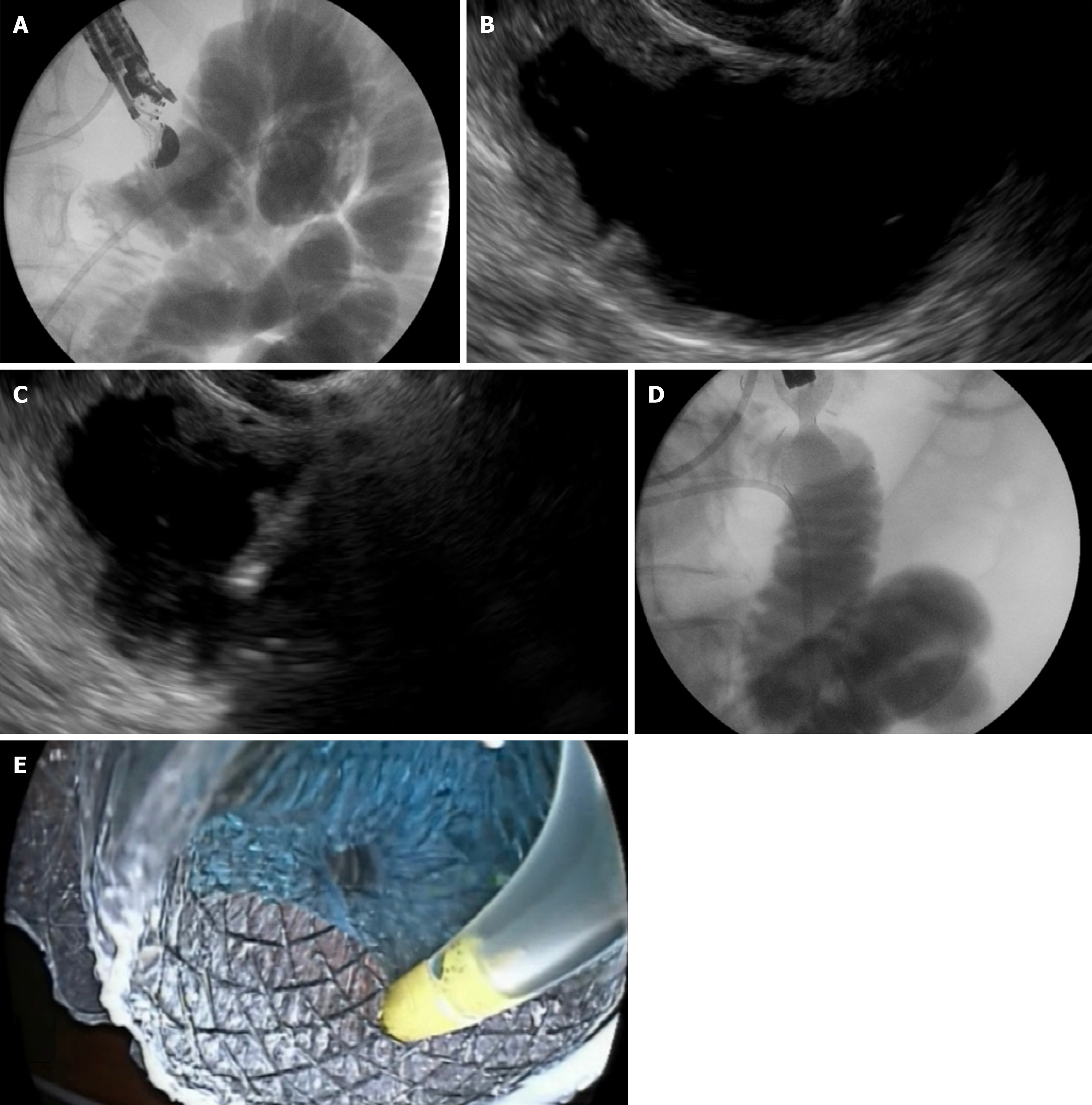
Figure 2 Step-by-step visualization of endoscopic ultrasound-guided gastroenterostomy procedure.
A: Fluoroscopic view showing a 7Fr nasobiliary catheter placed into the jejunum over a guidewire; B: Echoendoscopic view displaying the duodenum and jejunum distended with contrast material and methylene blue before direct puncture; C: Real-time puncture of the stomach and jejunal wall using the freehand technique under endoscopic ultrasound guidance; D: Fluoroscopic confirmation of successful stent deployment; E: Endoscopic confirmation ensuring correct positioning and lumen patency of the deployed stent.
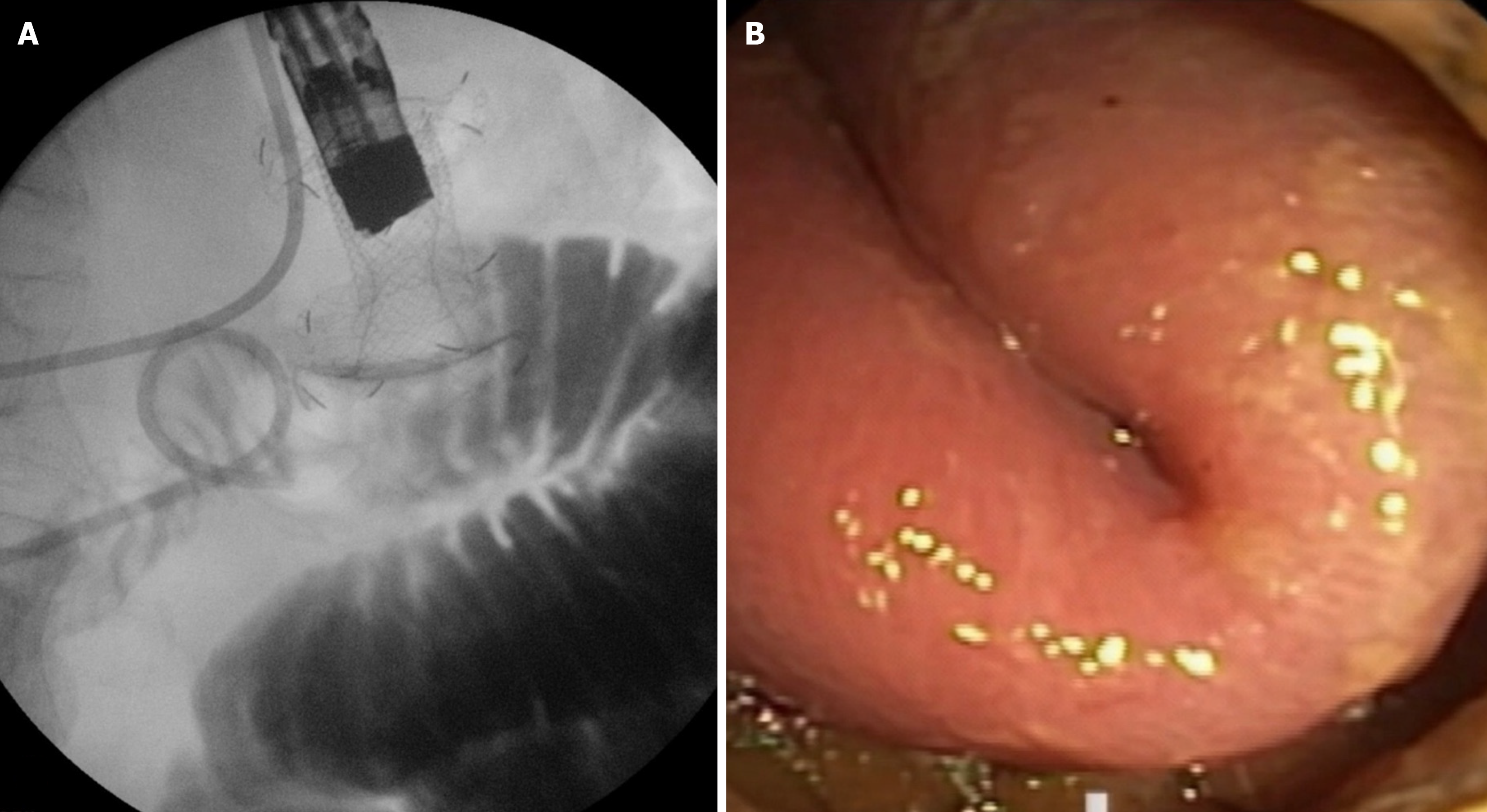
Figure 3 Type I stent misdeployment.
A: Fluoroscopic view showing the distal flange of the lumen-apposing metal stent deployed in the peritoneal cavity while the proximal flange remains in the stomach; B: Endoscopic view confirming the incomplete anastomosis with the small bowel intact.
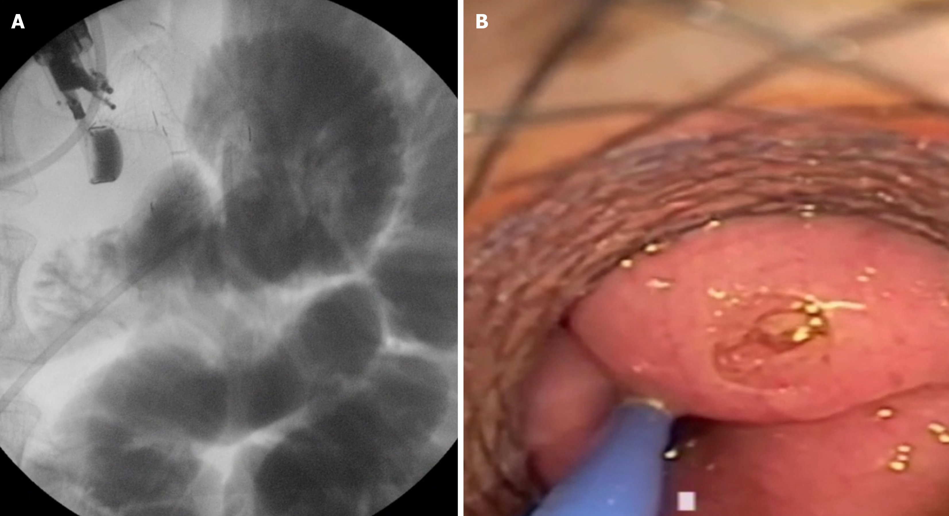
Figure 4 Type II stent misdeployment.
A: Fluoroscopic view demonstrating the distal flange of the lumen-apposing metallic stents deployed in the peritoneum with evidence of an enterotomy; B: Endoscopic view revealing the perforation at the small bowel site, requiring further intervention.
- Citation: Karagyozov PI, Kavrakov D, Shumka N. Endoscopic ultrasound-guided gastroenterostomy: The new standard treatment of gastric outlet obstruction. Artif Intell Gastrointest Endosc 2025; 6(2): 106600
- URL: https://www.wjgnet.com/2689-7164/full/v6/i2/106600.htm
- DOI: https://dx.doi.org/10.37126/aige.v6.i2.106600


