Copyright
©The Author(s) 2021.
Artif Intell Gastrointest Endosc. Aug 28, 2021; 2(4): 117-126
Published online Aug 28, 2021. doi: 10.37126/aige.v2.i4.117
Published online Aug 28, 2021. doi: 10.37126/aige.v2.i4.117
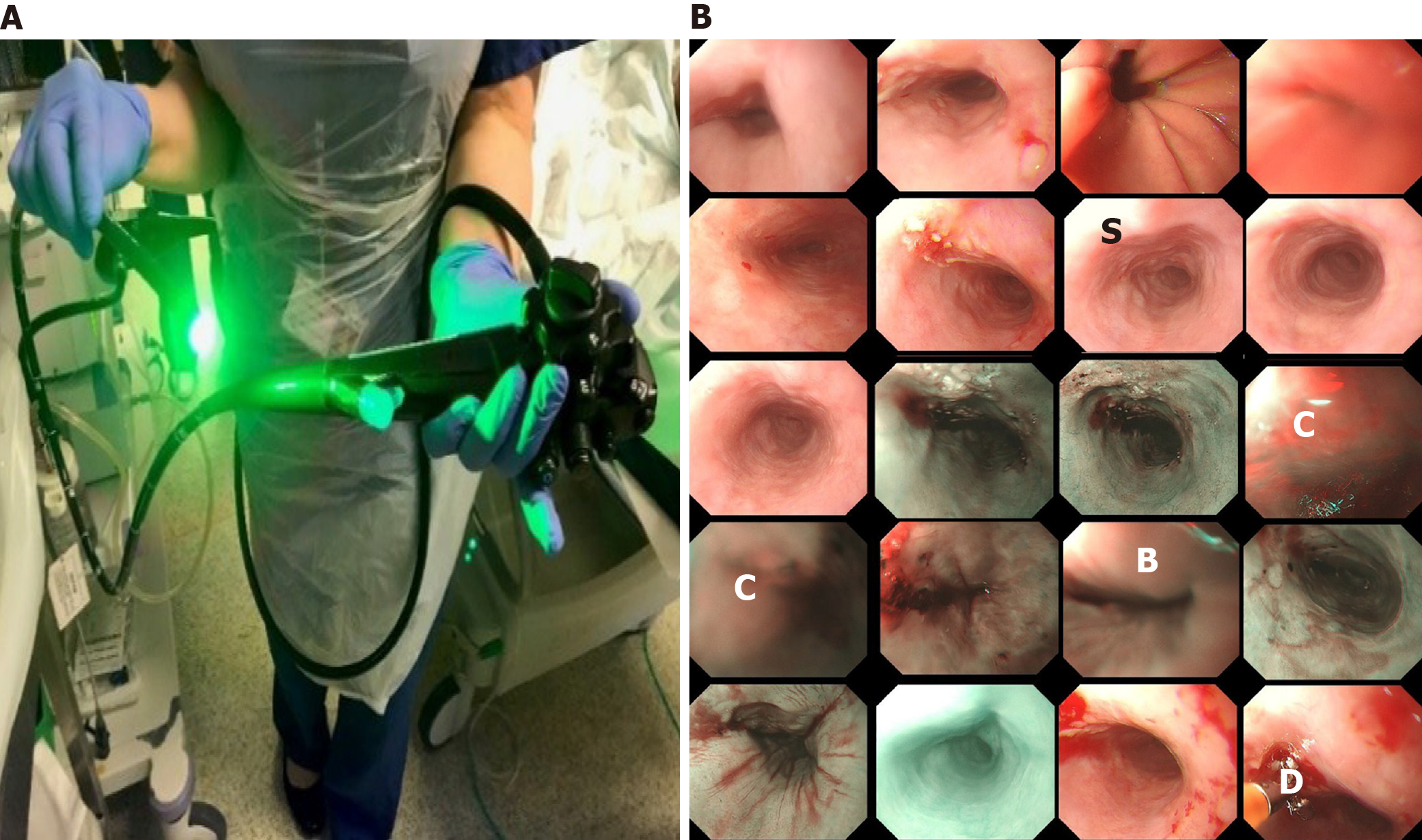
Figure 1 The endoscopy procedure.
A: The oesophagus camera; B: A montage display of a clip of an endoscopic video including narrow-band imaging and conventional white light endoscopy (e.g., top 2 rows). C: Colour misalignment; S: Saturation; B: Blurry; D: Device.
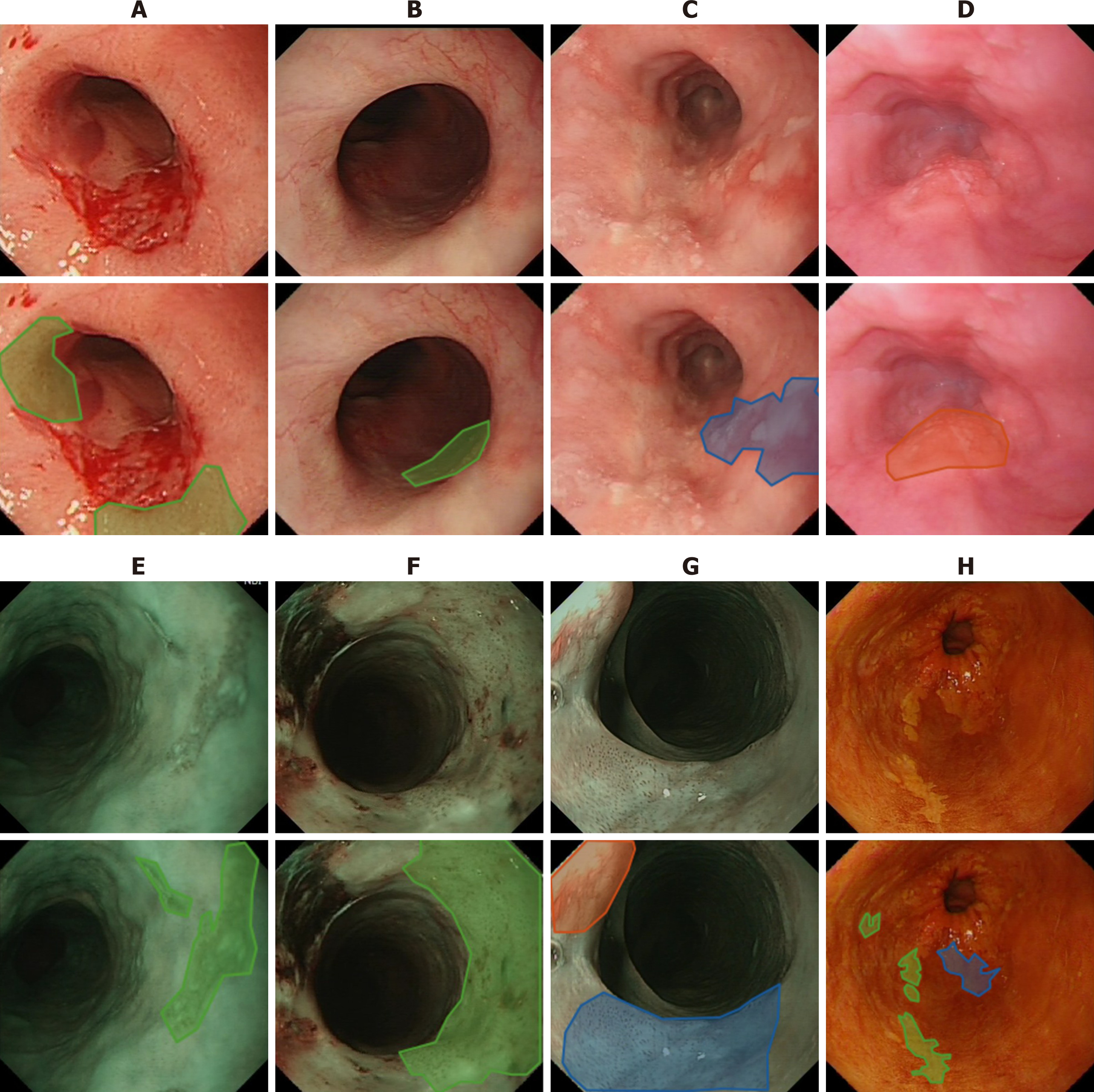
Figure 2 Examples of endoscopic images where green and blue masks refer to low and high grade dysplasia respectively and red for squamous cell cancer.
A-D: White light endoscopy; E-G: Narrow band imaging; H: Lugol’s. Mask colours: Green = low grade dysplasia; Blue = high grade dysplasia; Red = Squamous cell cancer.
- Citation: Gao X, Braden B. Artificial intelligence in endoscopy: The challenges and future directions. Artif Intell Gastrointest Endosc 2021; 2(4): 117-126
- URL: https://www.wjgnet.com/2689-7164/full/v2/i4/117.htm
- DOI: https://dx.doi.org/10.37126/aige.v2.i4.117


