Copyright
©The Author(s) 2020.
Artif Intell Med Imaging. Sep 28, 2020; 1(3): 94-107
Published online Sep 28, 2020. doi: 10.35711/aimi.v1.i3.94
Published online Sep 28, 2020. doi: 10.35711/aimi.v1.i3.94
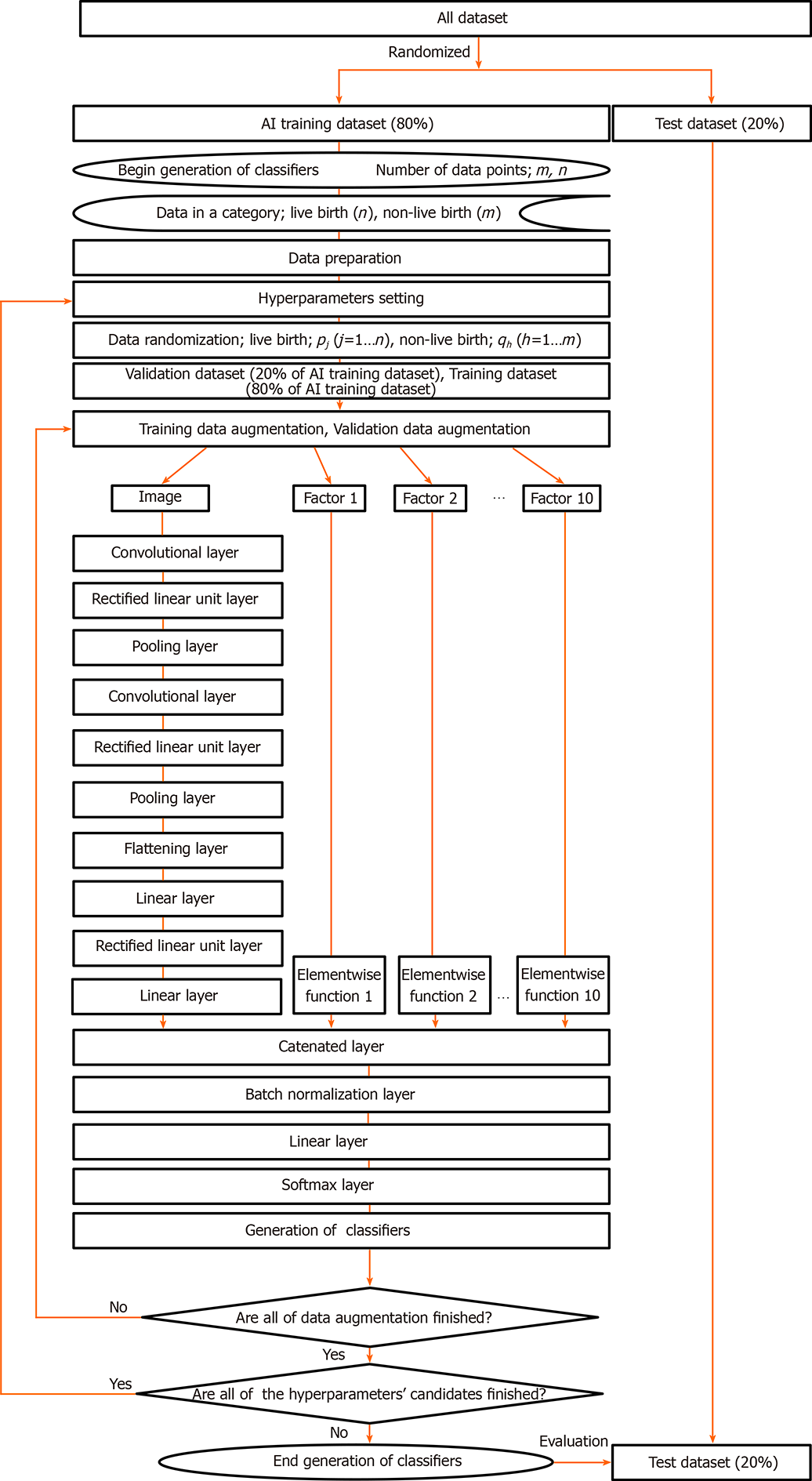
Figure 1 Flowchart for generating the artificial intelligence classifiers.
The artificial intelligence classifier consisted of a combination of 10 Layers of a convolutional neural network for an image and each elementwise layer often significant factors of the conventional embryo evaluation (CEE) that we had reported[14]. The ten factors chosen as independent factors to predict live birth were age, the number of embryo transfers, anti-Müllerian hormone concentration, day-3 blastomere number, grade on day 3, embryo cryopreservation day, inner cell mass, trophectoderm, average diameter, and body mass index. The functions in the elementwise layer for each factor of the CEE are shown as formulas in Table 1. The image processing and ten factors of the conventional embryo evaluation tensor were combined at the catenated layer. AI: Artificial intelligence.
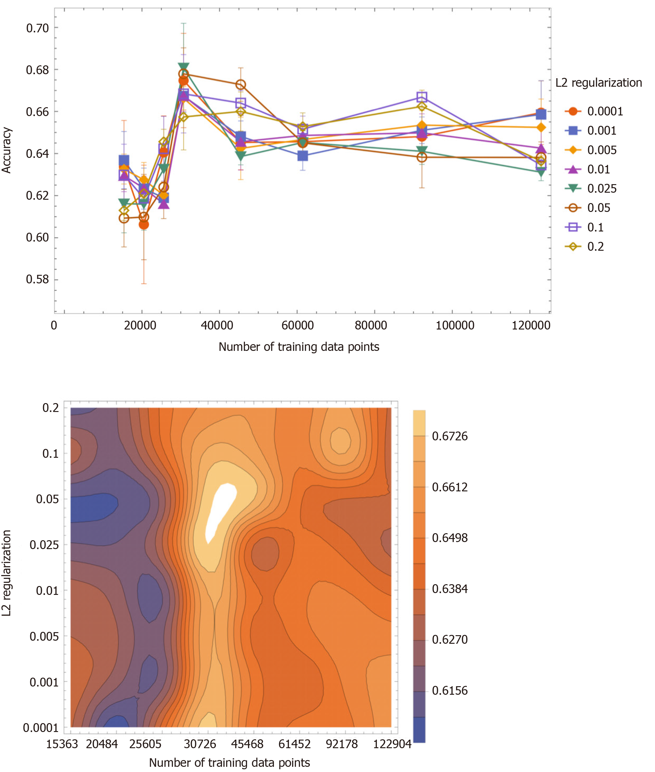
Figure 2 The accuracy value (mean ± SD) as a function of the number of training data points.
The accuracy values were classified with L2 regularization values: 0.0001, 0.001, 0.005, 0.01, 0.025, 0.05, 0.1 and 0.2. High accuracy values were obtained when the number of training data was between 25605 and 45468 (left panel). The two-dimensional contour plot of the accuracy value as a function of the training data and L2 regularization values (right panel). The brighter area indicates higher accuracy. High accuracy values were observed when the number of the training data was between 25605 and 45468 and when L2 regularization values were less than 0.1.
- Citation: Miyagi Y, Habara T, Hirata R, Hayashi N. Predicting a live birth by artificial intelligence incorporating both the blastocyst image and conventional embryo evaluation parameters. Artif Intell Med Imaging 2020; 1(3): 94-107
- URL: https://www.wjgnet.com/2644-3260/full/v1/i3/94.htm
- DOI: https://dx.doi.org/10.35711/aimi.v1.i3.94


