Copyright
©The Author(s) 2020.
Artif Intell Gastroenterol. Jul 28, 2020; 1(1): 19-29
Published online Jul 28, 2020. doi: 10.35712/aig.v1.i1.19
Published online Jul 28, 2020. doi: 10.35712/aig.v1.i1.19
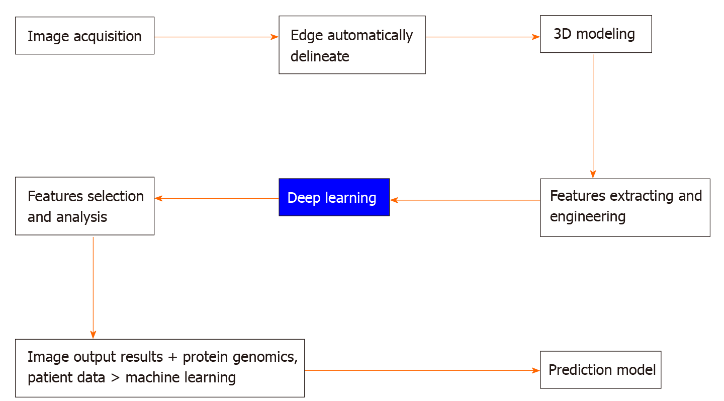
Figure 3 The entire process.
Machine learning is used to extract the imaging features of these cystic lesions, select and classify those features, and then use them to predict benign or malignant pancreatic cystic lesions. In this process, first, image acquisition is conducted uniformly, the edge of the suspected lesion object is delineated, and the three-dimensional shape of the variant is obtained. Then, the features of suspected diseases in the image are extracted, including the structure, density, and shape. AI software is used for in-depth learning, the features are screened and analyzed, and the imaging output results are obtained. The obtained results, proteomics, and patient data are entered into the machine learning model as the input layer to generate a predictive model, which can help clinicians in the differential diagnosis of benign and malignant pancreatic cysts.
- Citation: Lin HM, Xue XF, Wang XG, Dang SC, Gu M. Application of artificial intelligence for the diagnosis, treatment, and prognosis of pancreatic cancer. Artif Intell Gastroenterol 2020; 1(1): 19-29
- URL: https://www.wjgnet.com/2644-3236/full/v1/i1/19.htm
- DOI: https://dx.doi.org/10.35712/aig.v1.i1.19


