
















Copyright: ©Author(s) 2026.
World J Meta-Anal. Sep 18, 2026; 14(3): 123961
Published online Sep 18, 2026. doi: 10.13105/wjma.123961
Published online Sep 18, 2026. doi: 10.13105/wjma.123961
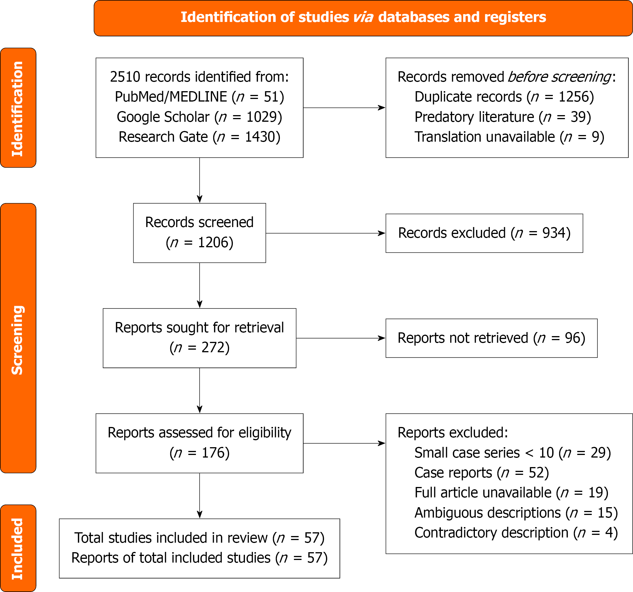
Figure 1
PRISMA flow diagram.
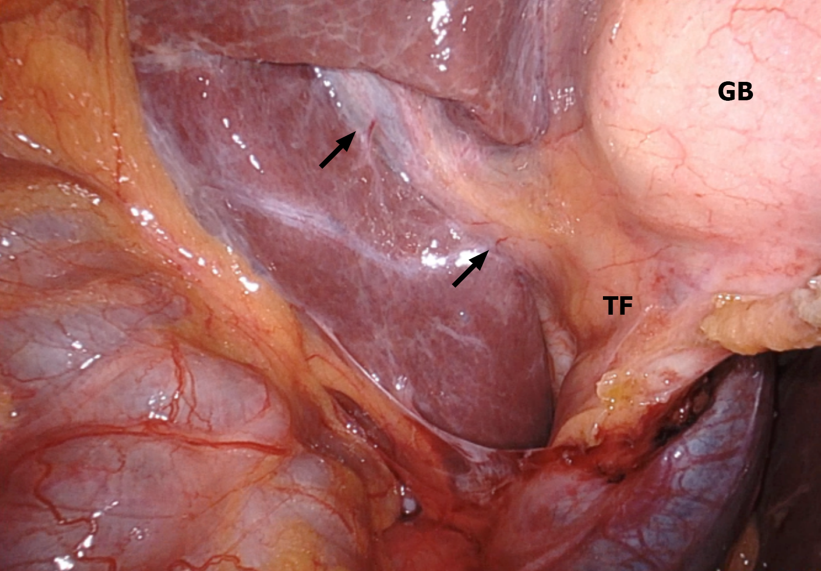
Figure 2 An intra-operative image during single incision laparoscopic cholecystectomy that demonstrates an open-type Rouvière’s sulcus (arrows), whose medial end is continuous with the transverse fissure, posterior to the gallbladder.
GB: Gallbladder; TF: Transverse fissure.
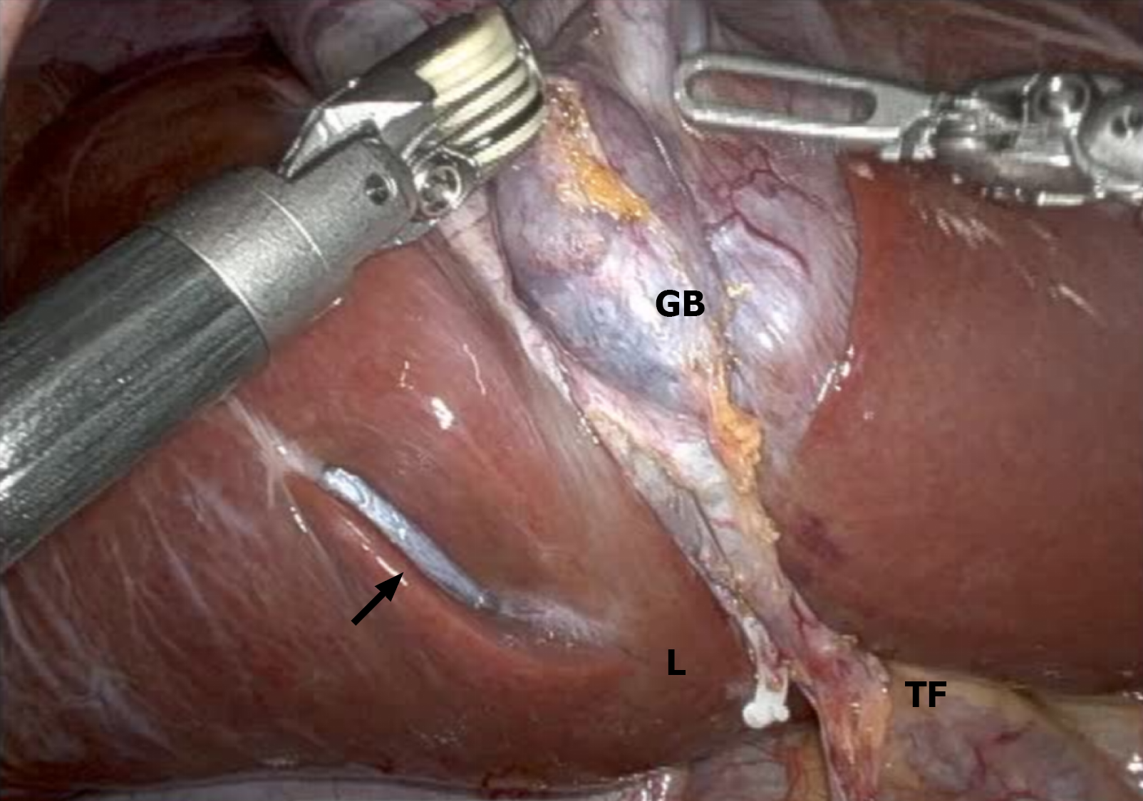
Figure 3 Intra-operative image during cholecystectomy, demonstrating a closed-type Rouvière’s sulcus, maintaining its posterior relationship to the gallbladder neck.
The medial end of Rouvière’s sulcus is separated from the transverse fissure by a bridge of liver parenchyma (L) so that the sulcus is not continuous with the transverse fissure. GB: Gallbladder; TF: Transverse fissure.
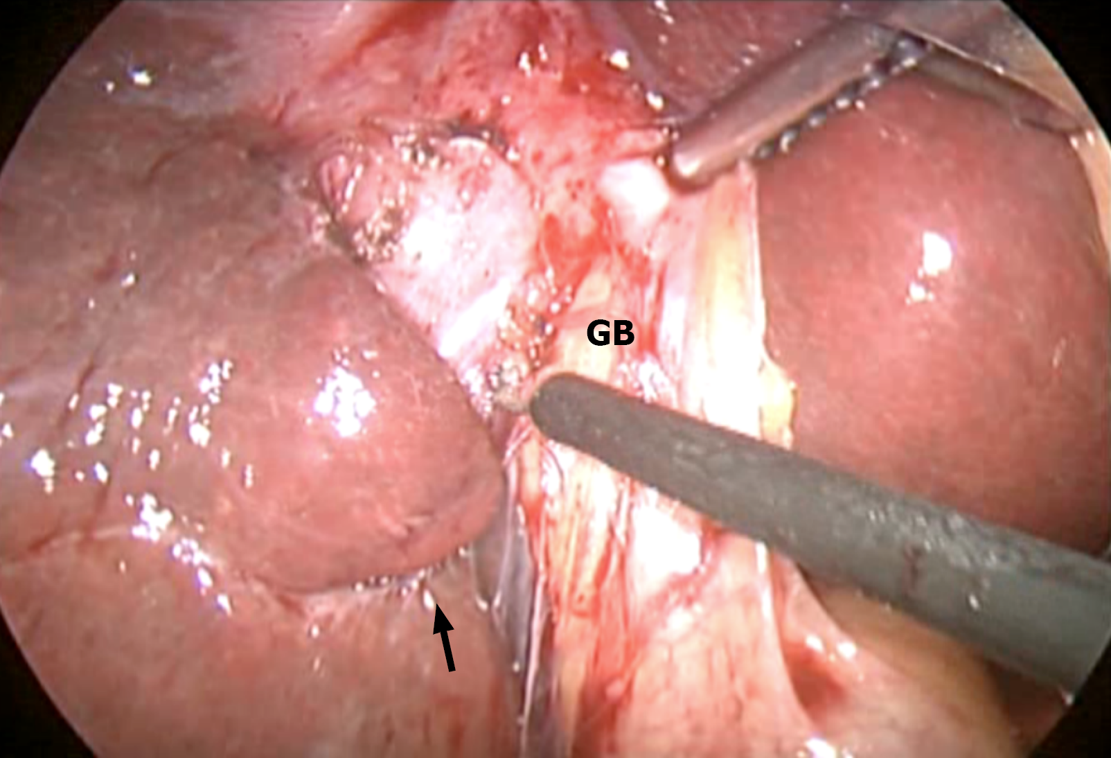
Figure 4 Intra-operative image taken during laparoscopic cholecystectomy, demonstrating a slit-type Rouvière’s sulcus (arrow), posterior to the gallbladder.
The sulcus is narrower than the operative instruments which are 5mm in diameter. GB: Gallbladder.
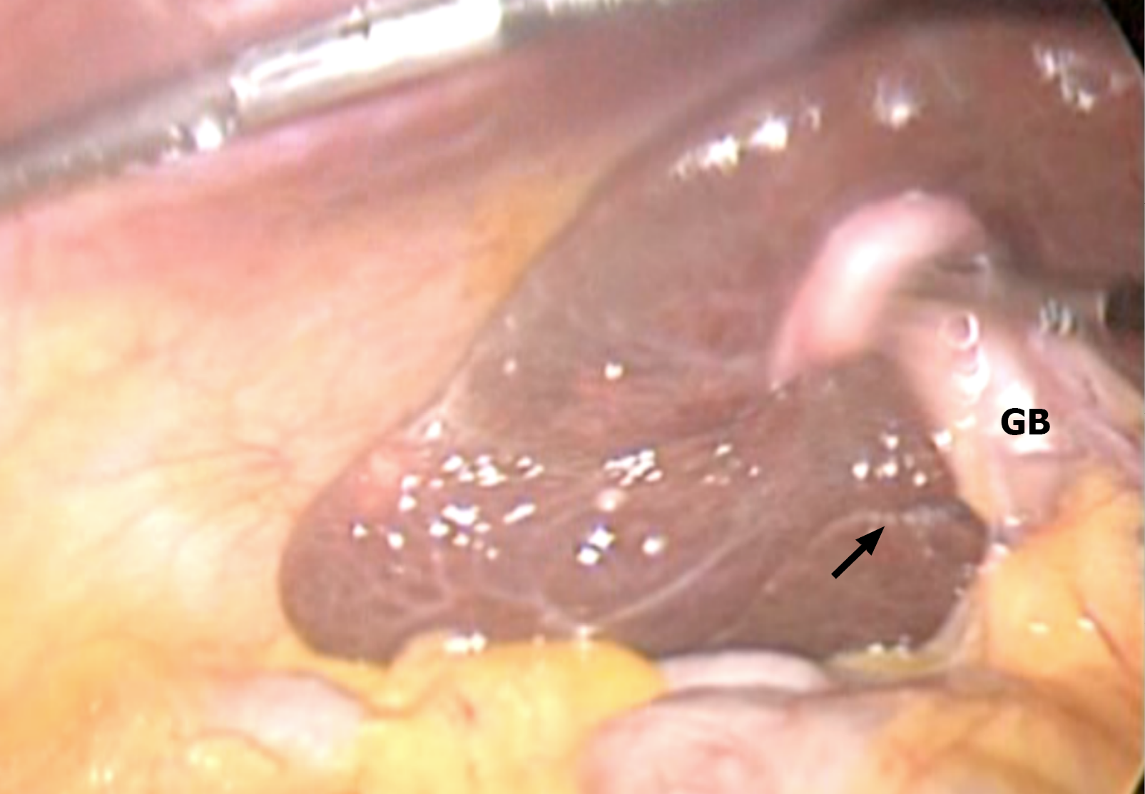
Figure 5 Intra-operative image taken at laparoscopic cholecystectomy, demonstrating a scar-type Rouvière’s sulcus (arrow).
There is only a vague impression/white line seen at the expected location of Rouvière’s sulcus, maintaining its posterior relationship to the gallbladder. GB: Gallbladder.
- Citation: Cawich SO, Kluger MD, Deshpande RR, Pearce NW, Alseidi A, Dapri G, Primrose JN, Gardner MT, Kedambady RS, Johnson S, Hewitt DB, Javed AA. Rouvière’s sulcus variants from a clinical perspective: A systematic review. World J Meta-Anal 2026; 14(3): 123961
- URL: https://www.wjgnet.com/2308-3840/full/v14/i3/123961.htm
- DOI: https://dx.doi.org/10.13105/wjma.123961