Copyright
©2013 Baishideng.
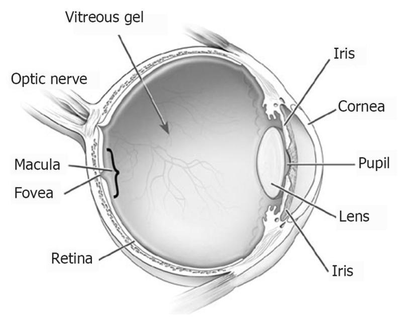
Figure 1 Structure of the eye.
Credit: National Eye Institute, National Institutes of Health.
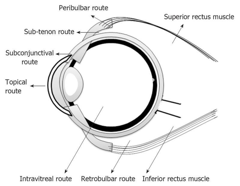
Figure 2 Routes of ocular drug delivery.
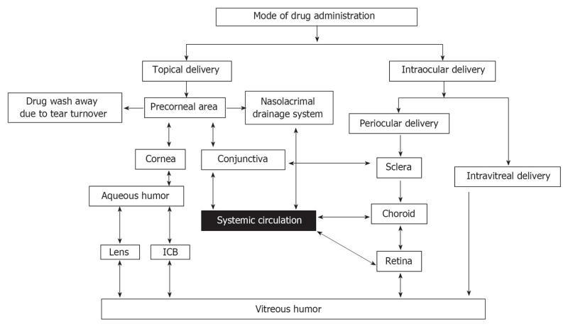
Figure 3 Disposition of drugs in the eye.
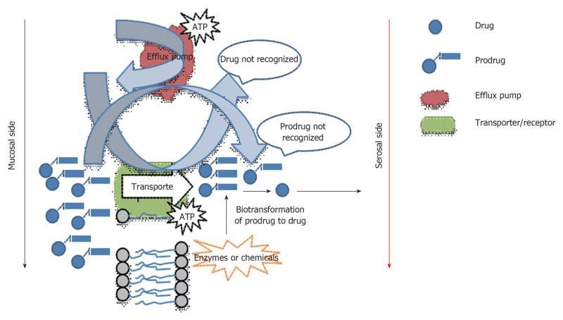
Figure 4 Role of efflux and influx transporters in ocular absorption.
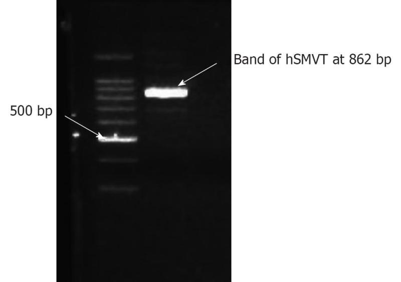
Figure 5 hSMVT cDNA was generated by reverse transcription polymerase chain reaction amplification of total RNA from ARPE-19 cells (lane 2).
Aliquots of polymerase chain reaction products were analyzed by gel electrophoresis on 0.8% agarose. Ethidium bromide staining of the gel showed a approximately 862 bp band corresponding. Reproduced with permission from[176].
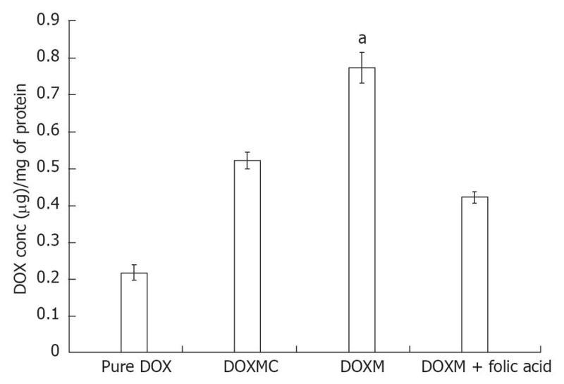
Figure 6 Quantitative uptake of doxorubicin in Y-79 cells using doxorubicin, doxorubicin-loaded in polymeric micelles, folate conjugated polymeric micelles and folate conjugated polymeric micelles in presence of folic acid.
aP < 0.05. Reproduced with permission from[186].
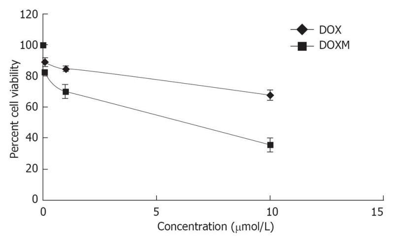
Figure 7 Cell viability studies of doxorubicin in ARPE-19 cells following treatment with doxorubicin and folate conjugated polymeric micelles.
Reproduced with permission from[186].
Figure 8 Reverse transcription polymerase chain reaction analysis of folate receptor-α, reduce folate carrier, proton coupled folate transporter.
GAPDH: Glyceraldehyde 3-phosphate dehydrogenase. Reproduced with permission from[180]. FR: Folate receptors; RFC: Reduced folate carrier; PCFT: Proton-coupled folate transporter.
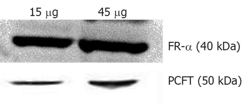
Figure 9 Western blotting analysis of folate receptor-α and proton coupled folate transporter.
Reproduced with permission from[180]. FR: Folate receptors; PCFT: Proton-coupled folate transporter.
- Citation: Boddu SH, Nesamony J. Utility of transporter/receptor(s) in drug delivery to the eye. World J Pharmacol 2013; 2(1): 1-17
- URL: https://www.wjgnet.com/2220-3192/full/v2/i1/1.htm
- DOI: https://dx.doi.org/10.5497/wjp.v2.i1.1


