
















Copyright: ©Author(s) 2026.
Artif Intell Gastrointest Endosc. Sep 8, 2026; 7(2): 122396
Published online Sep 8, 2026. doi: 10.37126/aige.122396
Published online Sep 8, 2026. doi: 10.37126/aige.122396
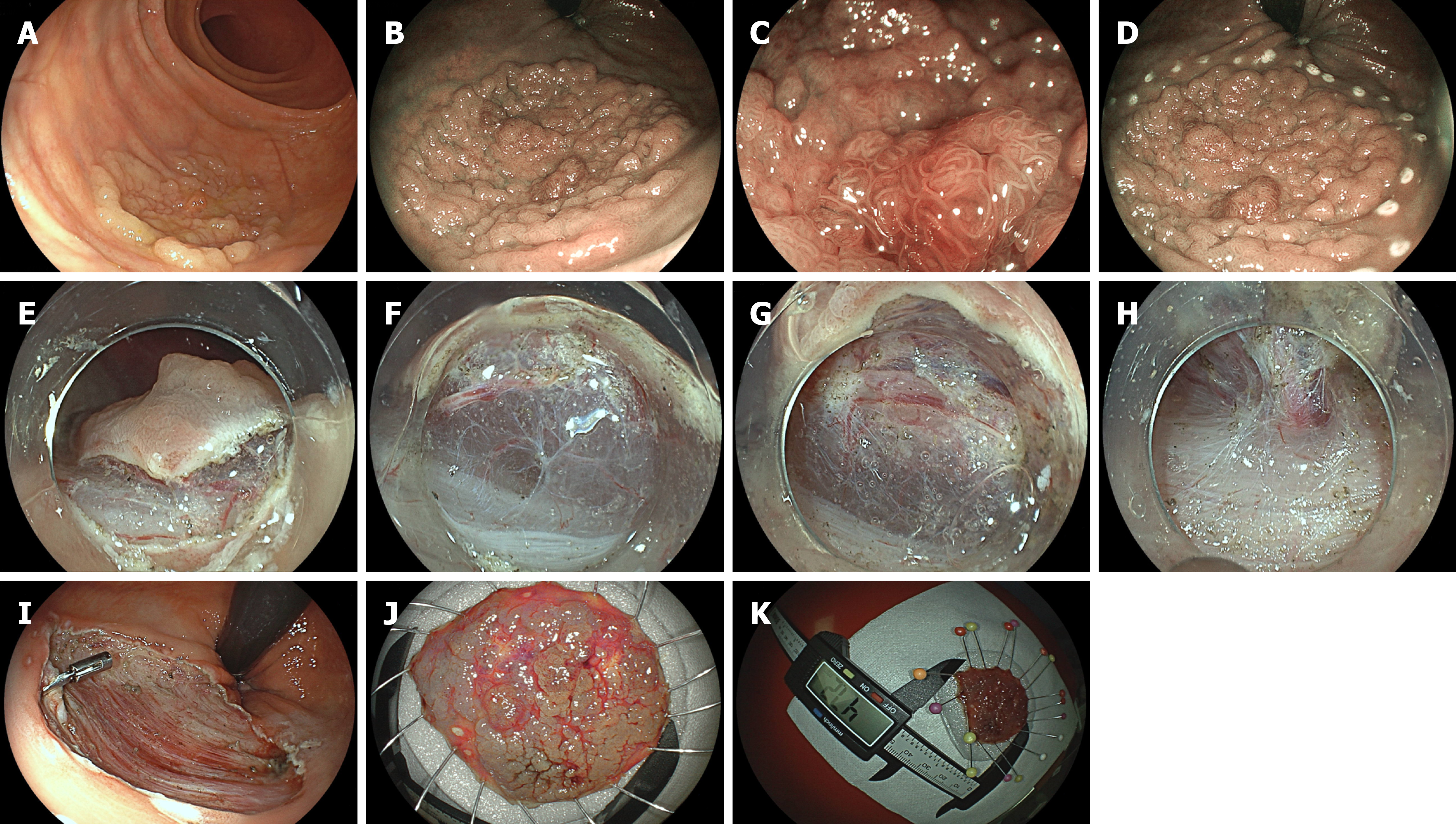
Figure 2 Representative case of colorectal endoscopic submucosal dissection.
A: Flat-elevated laterally spreading tumor in the rectum with a nodule in white-light imaging; B: Virtual chomoendoscopy assessment (Blue light imaging); C: Magnifying view assessing the nodule at the center of the lesion revealing enlarged crypts and slightly dilated and tortuous microvessels; D: Markings were placed with diathermic knife surrounding the lesion; E: Mucosal incision is performed in the anal side of the lesion; F: Pocket-creation method is initiated from anal side to oral side of the lesion; G: Pocket-creation method is advanced. Note that the dissection plane is at the deep submucosal layer, close to the muscularis propria layer; H: Perforating vessels should be identified, isolated and then coagulated with the knife or hemostatic forceps before transection; I: After endoscopic submucosal dissection is finished the defect should be closely inspected for blood vessel coagulation and closure of any muscularis propria layer damage with endoclips; J: The specimen should be fixed for histological assessment; K: The specimen measured 47.2 mm.
- Citation: Aliaga Ramos J, Nunes Arantes V. Colorectal endoscopic submucosal dissection up-to-date. Artif Intell Gastrointest Endosc 2026; 7(2): 122396
- URL: https://www.wjgnet.com/2689-7164/full/v7/i2/122396.htm
- DOI: https://dx.doi.org/10.37126/aige.122396