
















Copyright: ©Author(s) 2026.
Artif Intell Cancer. Sep 8, 2026; 7(1): 116460
Published online Sep 8, 2026. doi: 10.35713/aic.v7.i1.116460
Published online Sep 8, 2026. doi: 10.35713/aic.v7.i1.116460
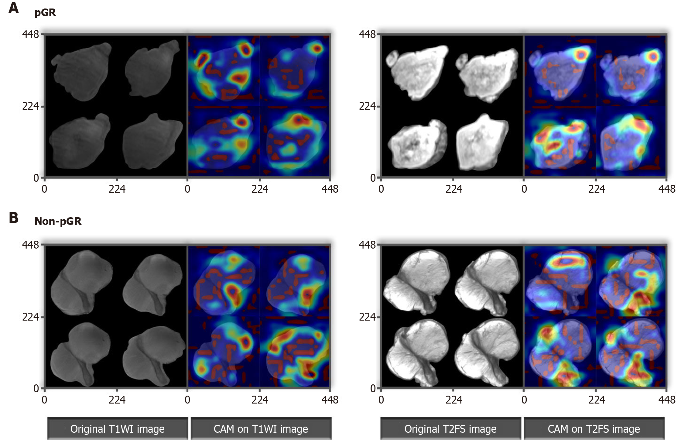
Figure 2 Feature heatmaps of representative patients on the deep learning ResNet50 algorithm via the Guided Gradient-weighted Class Activation Mapping in adolescents and young adults with osteosarcoma.
A: Pathological good response (pGR); B: Non-pGR. The original magnetic resonance imaging images and their corresponding feature heatmaps were shown from left to right. The red color highlighted the region of interest to classify pGR and non-pGR to neoadjuvant chemotherapy. The red color focused on different area for pGR (A) and non-pGR (B) on T1-weighted magnetic resonance imaging sequence (left) and fat-saturated T2-weighted magnetic resonance imaging sequence (right) magnetic resonance imaging images, respectively. The concentration of these heatmaps preferred central area for non-pGR images, and lesion boundaries for pGR images. pGR: Pathological good response; T1WI: T1-weighted magnetic resonance imaging sequence; CAM: Class activation mapping; T2FS: Fat-saturated T2-weighted magnetic resonance imaging sequence.
- Citation: Yang YH. Magnetic resonance imaging-based deep learning model for prediction of the neoadjuvant chemotherapy response and survival prognosis in adolescents with osteosarcoma. Artif Intell Cancer 2026; 7(1): 116460
- URL: https://www.wjgnet.com/2644-3228/full/v7/i1/116460.htm
- DOI: https://dx.doi.org/10.35713/aic.v7.i1.116460